Original Article – DOI: 10.33594/000000877
CPB (60): 376 - 385
Accepted: 20.07.2026 - Published: 29.07.2026
Adrenaline and Salbutamol Rapidly Exert Mast Cell-Stabilizing Properties in Rat Peritoneal Mast Cells
bTohoku Medical and Pharmaceutical University, Division of Respiratory Medicine, Sendai, Japan,
cYamagata University, School of Nursing, Yamagata, Japan
Keywords
Abstract
Background/Aims: Adrenaline is the first-choice drug for the treatment of anaphylaxis. Previous studies have revealed its prophylactic effects on the release of chemical mediators from mast cells. However, adrenaline quickly reverses the symptoms and signs of anaphylaxis after injection, indicating its rapid effects on mast cells. Methods: We examined the effects of adrenaline on rat peritoneal mast cell degranulation using differential-interference contrast (DIC) microscopy. To examine the rapid effects of adrenaline and salbutamol, a selective β2-adrenergic receptor agonist, we treated mast cells with these drugs immediately after exocytosis induction. Results: Similar to mast cells pre-incubated with adrenaline, those immediately treated with adrenaline did not exhibit exocytosis. At concentrations equal to or higher than 0.2 mg/ml, adrenaline significantly reduced the numbers of degranulating mast cells in a dose-dependent manner, whereas salbutamol required higher doses to exert significant effects. At 1 mg/ml, adrenaline and salbutamol almost completely suppressed mast cell degranulation. Conclusion: This study provides novel in vitro evidence indicating for the first time that adrenaline rapidly exerts mast cell-stabilizing properties in a dose-dependent manner. Salbutamol also stabilizes mast cells at high doses, revealing its additive therapeutic efficacy in anaphylaxis management.
Introduction
Anaphylaxis is an intense allergic response that can be life-threatening and is characterized by an acute syndrome affecting multiple systems owing to the rapid release of mediators from mast cells [1]. In the treatment, adrenaline, a non-specific adrenergic receptor agonist, is the first-choice drug, as it rapidly improves systemic circulation by inducing vasoconstriction and increasing blood pressure [2, 3]. In addition, adrenaline fundamentally prevents allergic reactions by suppressing the further release of chemical mediators from mast cells [2, 3]. Mechanistically, β2-adrenergic receptors are considered to play a key role, as activating these receptors significantly suppresses calcium mobilization in cells, which is dependent on high-affinity receptors for IgE (FceRI) [4]. Previous in vitro studies have revealed the prophylactic effects of adrenaline on mast cell degranulation by pre-incubating mast cells with adrenaline before inducing exocytosis [5-8]. However, in emergency situations, adrenaline quickly reverses the symptoms or signs of anaphylaxis after injection [2, 3]. This strongly indicates that adrenaline rapidly inhibits the release of chemical mediators from mast cells. In this study, to examine the rapid effects of adrenaline on stabilizing mast cells, we treated rat peritoneal mast cells with adrenaline immediately after exocytosis was induced. In addition, we analyzed the effects of salbutamol, a selective β2-adrenergic receptor agonist used to ameliorate bronchospasm caused by anaphylaxis [9]. For the first time, this study provides novel in vitro evidence showing that adrenaline rapidly exerts mast cell-stabilizing properties in a dose-dependent manner. Salbutamol also stabilizes mast cells at high doses, indicating its additive therapeutic efficacy in anaphylaxis management.
Materials and Methods
Cell Sources and Preparation
Male Wistar rats (aged a minimum of 25 weeks) were acquired from Jackson Laboratory Japan, Inc. (Yokohama, Japan). The rats were anesthetized using isoflurane and euthanized by cervical dislocation in accordance with the euthanasia guidelines for adult laboratory rodents [10]. The Animal Care and Use Committee of Miyagi University approved the animal protocol (No. 2026-02). As previously outlined [7, 11-19], the rat peritoneum was rinsed with a standard external (bathing) solution, which included: NaCl, 145 mM; KCl, 4.0 mM; CaCl2, 1.0 mM; MgCl2, 2.0 mM; HEPES, 5.0 mM; bovine serum albumin, 0.01 % (pH 7.2 adjusted with NaOH); and mast cells were isolated from the peritoneal cavity. The isolated mast cells were stored in the external solution at room temperature (22-24°C) for approximately 8 h until use. The mast cell suspension (approximately 200/µL) was distributed in a chamber positioned at the head stage of an inverted microscope (Nikon, Tokyo, Japan). Mast cells were easily identifiable from other cell types because of their distinctive intracellular secretory granules [7, 11-20]. Mast cell viability was assessed based on their ability to release secretory granules in response to external stimuli and their morphological integrity using differential-interference contrast (DIC) microscopy, as previously described [19, 21, 22].
Quantification of Mast Cell Degranulation
Adrenaline was purchased from Daiichi Sankyo, Inc. (Tokyo, Japan). As previously described [7, 19], mast cells were pre-incubated with 1 mg/ml (original concentration) adrenaline for 10 min, and exocytosis was externally induced by compound 48/80 (Sigma-Aldrich Co., St. Louis, MO, USA; final concentration, 10 µg/mL) (Fig. 1A top). To examine the rapid effects of adrenaline on mast cell degranulation and its therapeutic effects on degranulated cells, mast cells were treated with 1mg/ml adrenaline immediately (Fig. 1A middle) or 2 min (Fig. 1A bottom) after exocytosis induction by compound 48/80. To compare the rapid effects of adrenaline with those of salbutamol, purchased from GlaxoSmithKline K.K. (Tokyo, Japan), the drugs were separately dissolved in the external solution at final concentrations of 0.1, 0.2, 0.5, and 1 mg/ml (adrenaline, 0.55, 1.1, 2.7, and 5.5 mM; salbutamol, 0.17, 0.35, 0.87, and 1.7 mM). Mast cells were treated with these solutions or a solution without drugs immediately after exocytosis induction by compound 48/80 (Fig. 1B). Bright-field images were captured from randomly selected 0.1-mm2 fields of view (six to eight views from each condition), as previously described [7, 11-20]. The number of degranulated mast cells (defined as cells surrounded by more than eight granules outside the cell membrane) were counted and their ratio to the total number of mast cells was calculated.
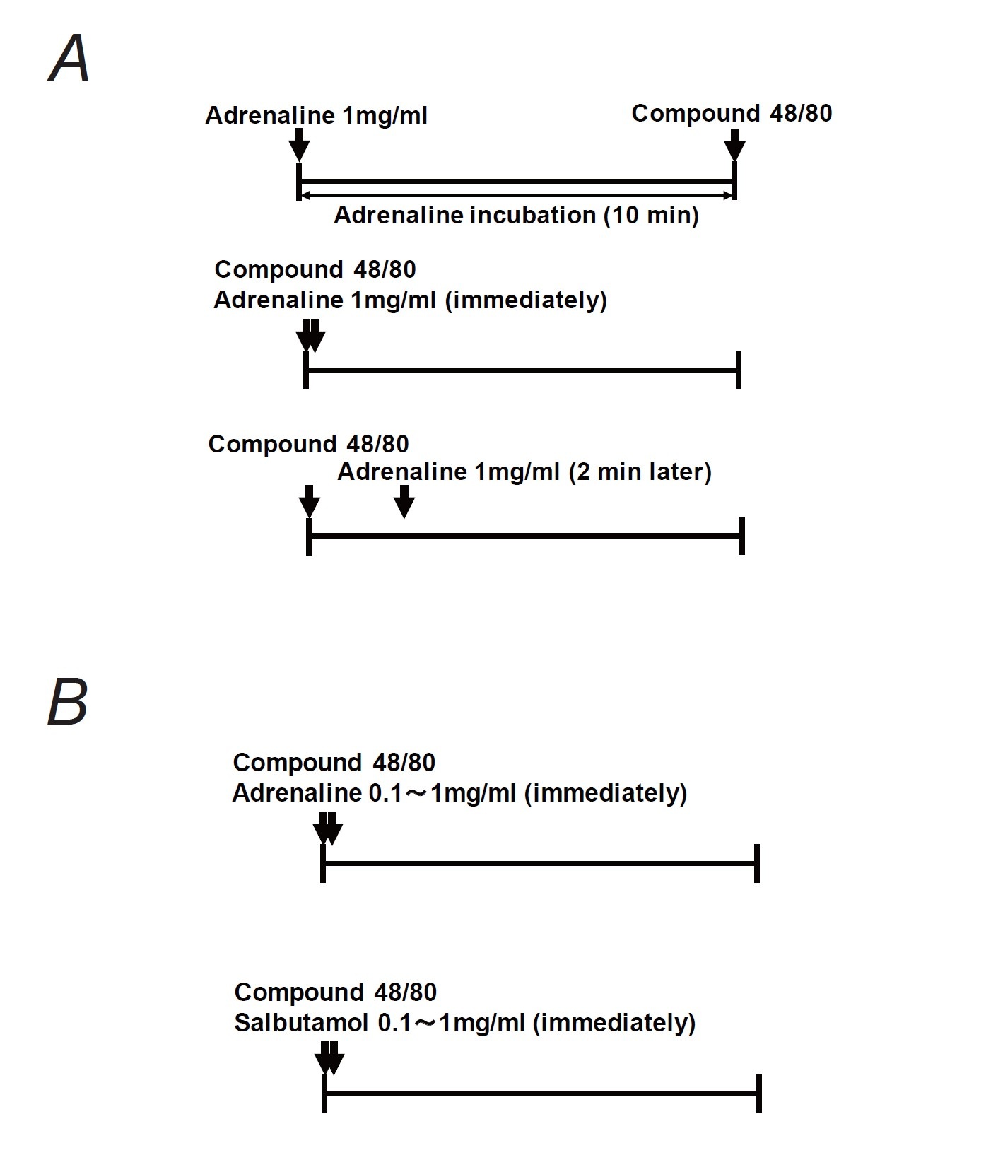
Fig. 1: Experimental protocols. A: Mast cells were pre-incubated with 1mg/ml adrenaline for 10 min and exocytosis was externally induced by compound 48/80 (top). To examine the rapid effects of adrenaline on mast cell degranulation and its therapeutic effects on degranulated cells, mast cells were treated with 1 mg/ml adrenaline immediately (middle) or 2 min after (bottom) exocytosis induction by compound 48/80. B: To compare the effects of adrenaline with those of salbutamol, mast cells were treated with adrenaline (top)- or salbutamol (bottom)-containing solutions (0.1, 0.2, 0.5, or 1 mg/ml) immediately after exocytosis induction by compound 48/80.
Statistical Analysis
Data were analyzed using Microsoft Excel (Microsoft Corporation, Redmond, WA., USA) and reported as means ± standard error of the mean (SEM). Statistical significance was assessed using analysis of variance (ANOVA) and set at p < 0.05.
Results
Effects of adrenaline on mast cell degranulation
When mast cells were exposed to compound 48/80, their surfaces became heavily wrinkled and the secretory granules were discharged through exocytosis (Fig. 2Ab vs. a). Consistent with our previous findings [7], mast cells pre-incubated with 1mg/ml adrenaline did not exhibit any of these characteristics (Fig. 2Ac) and the number of degranulating cells was markedly suppressed (compound 48/80, 100 ± 0.00 % vs. adrenaline (10 min) + compound 48/80, 12.1 ± 5.45 %; n=6, P<0.05; Fig. 2B). These results confirmed our previous findings that pre-incubation with adrenaline strongly inhibits the degranulation of mast cells and thus exerts mast cell-stabilizing properties [7]. To examine the rapid effects of adrenaline on the stabilization of mast cells and its therapeutic effects on degranulated cells, mast cells were treated with 1mg/ml adrenaline immediately or 2 min after exocytosis induction by compound 48/80. In mast cells treated with adrenaline immediately after exocytosis induction (Fig. 2Ad), similar to those after pre-incubation (Fig. 2Ac), the findings of exocytosis were almost completely absent and the number of degranulating cells was almost entirely suppressed (5.89 ± 1.35 %; n=6, P<0.05; Fig. 2B). In contrast, in mast cells treated with adrenaline 2 min after exocytosis induction (Fig. 2Ad), the findings of exocytosis were maintained and the number of degranulating cells was comparable to those treated with compound 48/80 alone (98.6 ± 1.39 %; n=6; Fig. 2B). These findings revealed that adrenaline rapidly exerts mast cell-stabilizing properties in the absence of pre-incubation. However, adrenaline did not exert any therapeutic effects on degranulated cells.
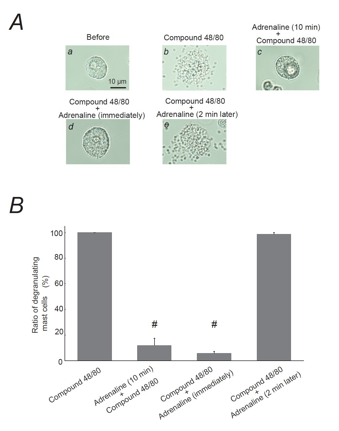
Fig. 2: Effects of adrenaline on mast cell degranulation. A: Differential-interference contrast (DIC) microscopic images were captured before (a) and after exocytosis was externally induced by compound 48/80 in mast cells (b-e). Mast cells were treated with compound 48/80 alone (b) or incubated with 1 mg/ml adrenaline before exocytosis induction (c). Other mast cells were treated with 1 mg/ml adrenaline immediately (d) or 2 min after (e) exocytosis induction. B: Mast cells were treated with compound 48/80 alone or incubated with 1 mg/ml adrenaline before exocytosis induction by compound 48/80. Other mast cells were treated with 1mg/ml adrenaline immediately or 2 min after exocytosis induction. Several samples of mast cell suspensions were obtained from the peritoneal cavity of a single rat. An aliquot of each sample was spread in a chamber placed at the head stage of an inverted microscope. Bright-field images were obtained from randomly selected 0.1-mm2 fields of view, in which 30-40 mast cells were evenly observed per field. Degranulating mast cells are expressed as the average percentage of total mast cells in the six bright fields. # p<0.05 vs. incubation in the external solution alone. Values were presented as the means ± standard error of the mean (SEM). Differences were analyzed using analysis of variance (ANOVA), followed by Dunnett’s t-test. The experiments were repeated at least thrice using three different rats to confirm the reproducibility of the data.
Rapid effects of adrenaline on mast cell degranulation
Based on our results, the original concentration of adrenaline (1 mg/ml) rapidly stabilized mast cells (Fig. 2Ad and B). Therefore, we further examined the rapid effects of adrenaline at lower concentrations (Fig. 3). Mast cells treated with the external solution alone or 0.1 mg/ml (0.55 mM) adrenaline showed the typical findings of exocytosis with a lot of wrinkles on their surface and secretory granules outside (Fig. 3Ab, c vs. a). However, these findings were partially or almost entirely absent in mast cells treated with higher concentrations (0.2, 0.5, and 1 mg/ml; 1.1, 2.7, and 5.5 mM) of adrenaline (Fig. 3Ad-f). Quantitatively, 0.1 mg/ml adrenaline did not affect the number of degranulating mast cells (Fig. 3B). However, 0.2 mg/ml adrenaline significantly decreased the number of degranulating mast cells (external solution, 100 ± 0.00 % vs. 0.2 mg/ml adrenaline, 73.4 ± 2.91 %; n=8, P<0.05) and 0.5 mg/ml showed more marked reduction (23.0 ± 3.08 %; n=7, P<0.05). One mg/ml adrenaline almost totally suppressed the number of degranulating mast cells (5.89 ± 1.35 %; n=6, P<0.05). These results indicate that, even in the absence of pre-incubation, adrenaline rapidly stabilized mast cells in a dose-dependent manner.
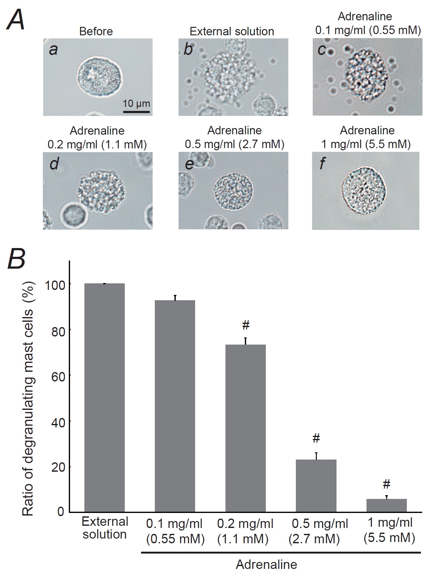
Fig. 3: Rapid effects of adrenaline on mast cell degranulation. A: Differential-interference contrast (DIC) microscopic images obtained before (a) and after exocytosis was externally induced by compound 48/80 in mast cells (b-f). Mast cells were immediately treated with the external solutions containing no substance (b) or 0.1 mg/ml (0.55 mM) adrenaline (c), 0.2 mg/ml (1.1 mM) adrenaline (d), 0.5 mg/ml (2.7 mM) adrenaline (e), or 1 mg/ml (5.5 mM) adrenaline (f). B: After exocytosis induction by compound 48/80, mast cells were immediately treated with the external solutions containing no substances or different concentrations of adrenaline. Several samples of mast cell suspensions were obtained from the peritoneal cavity of a single rat. An aliquot of each sample was spread in a chamber placed at the head stage of an inverted microscope. Bright-field images were obtained from randomly selected 0.1-mm2 fields of view, in which 30-40 mast cells were evenly observed per field. Degranulating mast cells are expressed as the average percentage of total mast cells in the six to eight bright fields. # p<0.05 vs. incubation in the external solution alone. Values were presented as the means ± SEM. Differences were analyzed using ANOVA followed by Dunnett’s t-test. The experiments were repeated at least thrice using three different rats to confirm the reproducibility of the data.
Rapid effects of salbutamol on mast cell degranulation
Adrenaline is a non-selective agonist of adrenergic receptors such as α1, α2, β1, and β2 [3]. Among these, the β2-adrenergic receptor transduces signals for adrenaline-induced inhibition of exocytosis in mast cells [4, 7]. Salbutamol is a selective, short-acting β2-agonist used to ameliorate bronchospasm caused by anaphylaxis [9]. Therefore, with the expectation of its β2-receptor mediated properties in mast cells, we also examined the rapid effects of salbutamol on the degranulation of the cells (Fig. 4). Relatively lower concentrations of salbutamol (0.1 and 0.2 mg/ml; 0.17 and 0.35 mM) did not affect the findings of exocytosis in mast cells (Fig. 4Ac, d vs. b) and the numbers of degranulating cells were almost comparable to those treated with the external solution alone (Fig. 4B). However, relatively higher concentrations of salbutamol (0.5 and 1 mg/ml; 0.87 and 1.7 mM) partially or entirely halted exocytosis (Fig. 4Ae and f). Quantitatively, 0.5 mg/ml salbutamol significantly reduced the number of degranulating mast cells (external solution, 99.7 ± 3.47 % vs. 0.5 mg/ml salbutamol, 79.0 ± 2.56 %; n=6, P<0.05; Fig. 4B). Notably, 1 mg/ml salbutamol almost totally suppressed the number of degranulating mast cells (8.14 ± 2.82 %; n=6, P<0.05). These results revealed that, similar to adrenaline (Fig. 3), salbutamol rapidly stabilized mast cells in a dose-dependent manner. From our results, the effects of salbutamol appeared to be less pronounced than those of adrenaline at all concentrations except for 1 mg/ml (Fig. 4B vs. 3B). However, when compared on a molar basis, salbutamol required lower concentrations than adrenaline to produce comparable effects (Fig. 4B vs. 3B). These findings indicate that salbutamol may exert more potent mast cell-stabilizing properties than adrenaline.
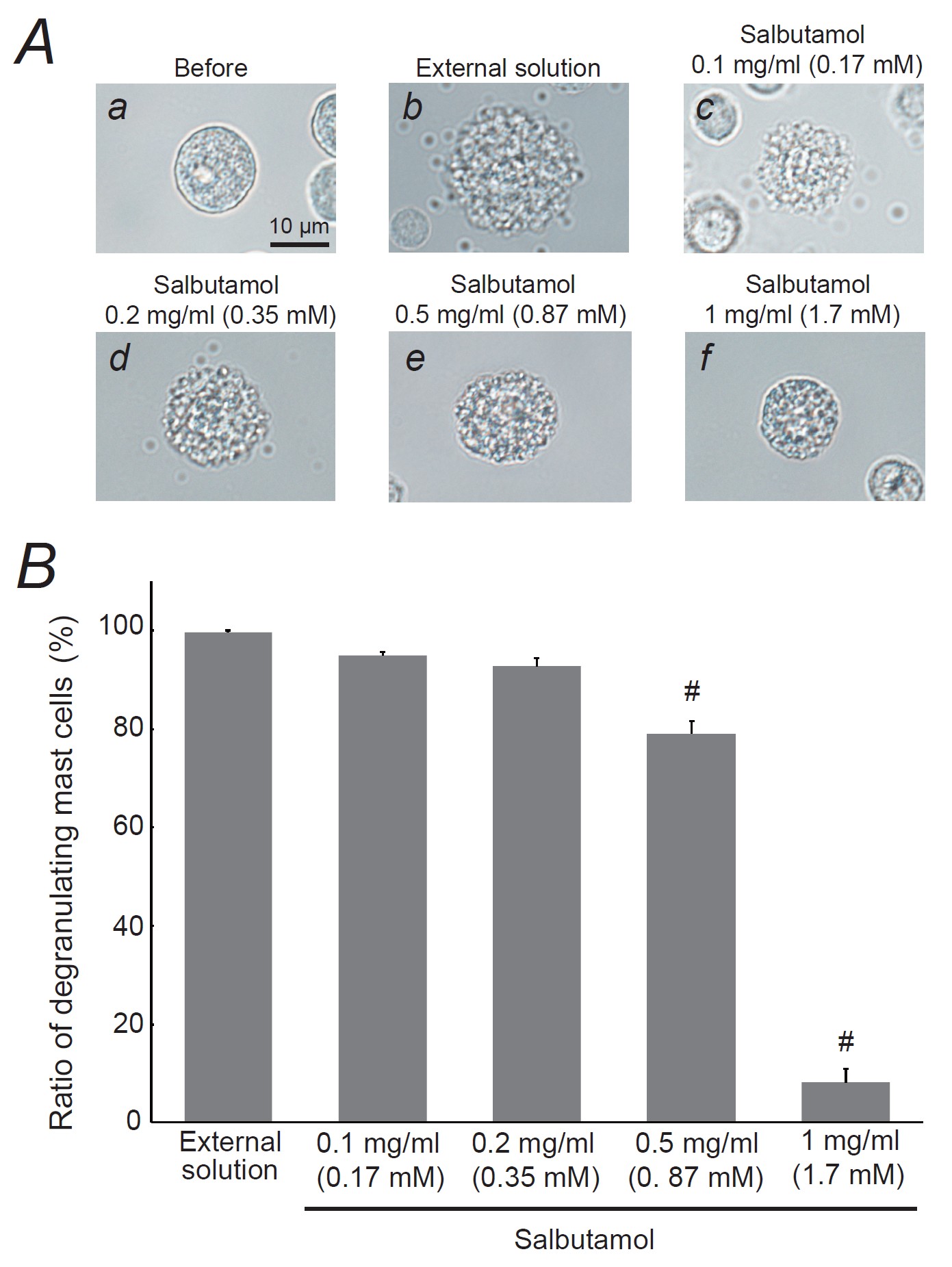
Fig. 4: Rapid effects of salbutamol on mast cell degranulation. A: Differential-interference contrast (DIC) microscopy images obtained before (a) and after exocytosis was externally induced by compound 48/80 in mast cells (b-f). Mast cells were immediately treated with the external solutions containing no substance (b) or 0.1 mg/ml (0.17 mM) salbutamol (c), 0.2 mg/ml (0.35 mM) salbutamol (d), 0.5 mg/ml (0.87 mM) salbutamol (e), and 1 mg/ml (1.7 mM) salbutamol (f). B: After exocytosis induction by compound 48/80, mast cells were immediately treated with the external solutions containing no substances or different concentrations of salbutamol. Several samples of mast cell suspensions were obtained from the peritoneal cavity of a single rat. An aliquot of each sample was spread in a chamber placed at the head stage of an inverted microscope. Bright-field images were obtained from randomly selected 0.1-mm2 fields of view, in which 30-40 mast cells were evenly observed per field. Degranulating mast cells are expressed as the average percentage of total mast cells in the six to eight bright fields. # p<0.05 vs. incubation in the external solution alone. Values were presented as the means ± SEM. Differences were analyzed using ANOVA followed by Dunnett’s t-test. The experiments were repeated at least thrice using three different rats to confirm the reproducibility of the data.
Discussion
Our studies have shown that adrenaline possesses properties that stabilize mast cells through the ongoing in vitro observation of exocytosis in these cells [7, 19]. In these studies, mast cells were pre-incubated with adrenaline before exocytosis induction. As antigen binding to IgE on mast cells triggers a rapid anaphylactic response, compound 48/80 prompts mast cell degranulation within 10 s of being introduced [23]. In the present study, to examine the rapid effect of adrenaline on the degranulation of mast cells, the cells were treated with adrenaline immediately after exocytosis induction (Fig. 1A). As shown in Figures 2Ae and B, adrenaline did not exert therapeutic effects by reversing the ongoing degranulation of mast cells. However, despite the absence of pre-incubation, adrenaline rapidly exerted prophylactic effects by suppressing the further initiation of exocytosis (Fig. 2Ac and B), and these effects were dose-dependent (Fig. 3). Adrenaline is the first-choice drug for anaphylaxis management because it rapidly improves systemic circulation by inducing vasoconstriction and increasing blood pressure [9]. The present study revealed the additive prophylactic effect of adrenaline on rapidly stabilizing mast cells immediately after administration (Fig. 5). Our results showed that salbutamol, a selective β2-adrenergic receptor agonist, also rapidly exerted mast cell-stabilizing effects in a dose-dependent manner (Fig. 4). Notably, at 1 mg/ml, salbutamol stabilized mast cells as potently as adrenaline. Additionally, when compared on a molar basis, salbutamol required lower concentrations than adrenaline to produce comparable effects (Fig. 4 vs. 3). Previous studies using mast cells isolated from human lung tissue have shown that β2-agonists, including salbutamol, terbutaline, and isoprenaline, effectively inhibited the IgE-dependent release of histamine [24, 25]. In addition to chemical mediators such as histamine, serotonin, leukotrienes, and prostaglandins, mast cells release various cytokines and growth factors through exocytosis [26]. Therefore, to accurately assess the mast cell-stabilizing effects of drugs or substances, it is more effective to directly observe exocytosis instead of indirectly measuring the levels of released chemical mediators [12, 13, 15, 27]. In this study, we microscopically examined the entire exocytosis process using mast cells extracted from rat peritoneal cavities and quantified the process by determining the proportion of degranulating mast cells. In anaphylaxis management, the use of salbutamol is recommended because it ameliorates bronchospasm through β2- adrenergic receptor stimulation [9]. Our current findings revealed the additive therapeutic efficacy of salbutamol as a rapid and potent mast cell stabilizer for anaphylaxis management (Fig. 5). However, the concentrations of adrenaline and salbutamol used in the present study are substantially higher than standard physiological levels [28, 29]. In clinical settings, the actual concentrations reaching the systemic circulation and tissues to interact with mast cells are significantly lower [28, 29]. Therefore, our findings should be interpreted with caution when considering their clinical application. Earlier research utilizing human lung mast cells demonstrated that the primary mechanism for the inhibition of exocytosis by β2-agonists involves the activation of β2-adrenergic receptors. This process is associated with calcium mobilization dependent on cyclic AMP, facilitated through the coupling of G-proteins [4, 30]. In our previous study, pre-incubation with adrenaline inhibited the induction of exocytosis in rat peritoneal mast cells [7], which was almost completely restored in the presence of butoxamine, a specific β2-adrenergic receptor antagonist [7]. This indicated the involvement of β2-adrenergic receptors in the adrenaline-induced inhibition of exocytosis. In the present study, to reveal the mechanisms underlying the mast cells-stabilizing properties of adrenaline and salbutamol, the use of ICI 118, 551, a highly selective β2-adrenergic receptor antagonist [31], or assessing intracellular signaling molecules such as cyclic AMP and calcium dynamics would be beneficial. However, the lack of such mechanistic data is a limitation of this study. In our previous study, we showed through continuous observation of exocytosis in mast cells that macrolide antibiotics such as clarithromycin, corticosteroids such as dexamethasone and hydrocortisone, anti-hypertensive drugs such as prazosin, and anti-allergic medications including tranilast, ketotifen, olopatadine, and cetirizine possess properties that stabilize mast cells [7, 12-15, 18]. Moreover, we showed that certain food components, such as caffeine, catechins, and vitamins, along with compounds present in lemon juice or peel like citric acid, hesperetin, and eriodictyol, stabilize mast cells and have synergistic effects when used together [16, 17, 20]. Recently, using the same approach, we showed that essential trace elements, specifically magnesium and zinc, inhibit exocytosis in a dose-dependent manner, thus exhibiting mast cell-stabilizing effects [19]. From our results, both adrenaline and salbutamol rapidly exerted mast cell-stabilizing properties in a dose-dependent manner (Fig. 3 and 4). However, these effects were not enough at lower doses. In our recent study, we revealed that high levels of magnesium and zinc chloride amplified the stabilizing effects of adrenaline on mast cells [19]. Therefore, the administration of drugs or food constituents that stabilize mast cells may additively or synergistically enhance the rapid effects of adrenaline or salbutamol in anaphylaxis management.
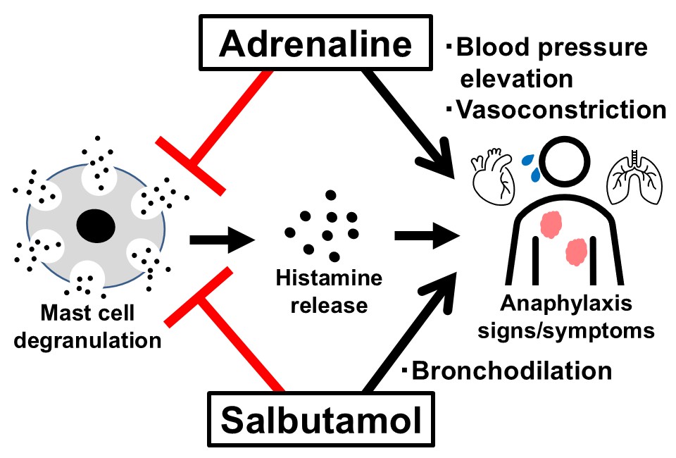
Fig. 5: Mast cell-stabilizing properties of adrenaline and salbutamol. Adrenaline is the first-choice drug for anaphylaxis management because it quickly ameliorates systemic circulation by causing vasoconstriction and elevating blood pressure. The present study revealed an additional property of adrenaline in rapidly stabilizing mast cells immediately after administration. However, the use of salbutamol is also recommended in anaphylaxis management because it ameliorates bronchospasm through β2- adrenergic receptor stimulation. Our findings present the additive therapeutic efficacy of salbutamol as a rapid and potent mast cell stabilizer for the anaphylaxis management.
Conclusion
For the first time, this study provides novel in vitro evidence showing that adrenaline rapidly exerts mast cell-stabilizing properties in a dose-dependent manner. Salbutamol, a selective β2-adrenergic receptor agonist, also stabilizes mast cells at high doses, indicating its additive therapeutic efficacy in anaphylaxis management. However, the concentrations of adrenaline and salbutamol used in the present study are substantially higher than standard physiological levels. Therefore, our findings should be interpreted with caution when considering their clinical application.
Acknowledgements
Special thanks We are grateful to Mr. Kanata Segawa, Ms. Aiko Abe, Ms. Michiko Shoji, and Ms. Kanako Sugawara for their constant encouragement and moral support.
Author contributions
HH, KS, KK, and HS performed the experiments and analyzed the data. TT and JS helped analyze the data. IK designed the experiments, interpreted the results and wrote the manuscript. All the authors have read and approved the final version of the manuscript for publication.
Funding
This study was supported by the Tojuro Iijima Foundation for Food Science and Technology (No. 2025-28 to IK).
Availability of data and materials
Data used to support the findings of this study are available from the corresponding author upon reasonable request.
Ethics approval and consent to participate
This study was performed in accordance with the Guide for the Care and Use of Laboratory Animals of Miyagi University, which includes ethical considerations.
Disclosure Statement
The authors declare no Disclosure Statement.
References
- Sampson HA, Munoz-Furlong A, Campbell RL, Adkinson NF, Jr., Bock SA, Branum A, Brown SG, Camargo CA, Jr., Cydulka R, Galli SJ, Gidudu J, Gruchalla RS, Harlor AD, Jr., Hepner DL, Lewis LM, Lieberman PL, Metcalfe DD, O'Connor R, Muraro A, Rudman A, et al.: Second symposium on the definition and management of anaphylaxis: summary report--Second National Institute of Allergy and Infectious Disease/Food Allergy and Anaphylaxis Network symposium. J Allergy Clin Immunol 2006;117:391-397. https://doi.org/10.1016/j.jaci.2005.12.1303
- Kemp SF, Lockey RF, Simons FE, World Allergy Organization ad hoc Committee on Epinephrine in A: Epinephrine: the drug of choice for anaphylaxis-a statement of the world allergy organization. World Allergy Organ J 2008;1:S18-26. https://doi.org/10.1186/1939-4551-1-S2-S18
- Ring J, Klimek L, Worm M: Adrenaline in the Acute Treatment of Anaphylaxis. Dtsch Arztebl Int 2018;115:528-534. https://doi.org/10.3238/arztebl.2018.0528
- Kuehn HS, Gilfillan AM: G protein-coupled receptors and the modification of FcepsilonRI-mediated mast cell activation. Immunol Lett 2007;113:59-69. https://doi.org/10.1016/j.imlet.2007.08.007
- Graevskaya EE, Akhalaya MY, Goncharenko EN: Effects of cold stress and epinephrine on degranulation of peritoneal mast cells in rats. Bull Exp Biol Med 2001;131:333-335. https://doi.org/10.1023/A:1017991817000
- Moroni F, Fantozzi R, Masini E, Mannaioni PF: The modulation of histamine release by alpha-adrenoceptors: evidences in murine neoplastic mast cells. Agents Actions 1977;7:57-61. https://doi.org/10.1007/BF01964881
- Abe N, Toyama H, Ejima Y, Saito K, Tamada T, Yamauchi M, Kazama I: Prazosin Potentiates Mast Cell-Stabilizing Property of Adrenaline. Cell Physiol Biochem 2024;58:212-225. https://doi.org/10.33594/000000703
- Kazama I: Potential prophylactic efficacy of mast cell stabilizers against COVID-19 vaccine-induced anaphylaxis. Clin Mol Allergy 2021;19:25. https://doi.org/10.1186/s12948-021-00162-9
- Ring J, Beyer K, Biedermann T, Bircher A, Duda D, Fischer J, Friedrichs F, Fuchs T, Gieler U, Jakob T, Klimek L, Lange L, Merk HF, Niggemann B, Pfaar O, Przybilla B, Rueff F, Rietschel E, Schnadt S, Seifert R, et al.: Guideline for acute therapy and management of anaphylaxis: S2 Guideline of the German Society for Allergology and Clinical Immunology (DGAKI), the Association of German Allergologists (AeDA), the Society of Pediatric Allergy and Environmental Medicine (GPA), the German Academy of Allergology and Environmental Medicine (DAAU), the German Professional Association of Pediatricians (BVKJ), the Austrian Society for Allergology and Immunology (OGAI), the Swiss Society for Allergy and Immunology (SGAI), the German Society of Anaesthesiology and Intensive Care Medicine (DGAI), the German Society of Pharmacology (DGP), the German Society for Psychosomatic Medicine (DGPM), the German Working Group of Anaphylaxis Training and Education (AGATE) and the patient organization German Allergy and Asthma Association (DAAB). Allergo J Int 2014;23:96-112. https://doi.org/10.1007/s40629-014-0009-1
- Clarkson JM, Martin JE, McKeegan DEF: A review of methods used to kill laboratory rodents: issues and opportunities. Lab Anim 2022;56:419-436. https://doi.org/10.1177/00236772221097472
- Kazama I, Maruyama Y, Takahashi S, Kokumai T: Amphipaths differentially modulate membrane surface deformation in rat peritoneal mast cells during exocytosis. Cell Physiol Biochem 2013;31:592-600. https://doi.org/10.1159/000350079
- Baba A, Tachi M, Maruyama Y, Kazama I: Olopatadine inhibits exocytosis in rat peritoneal mast cells by counteracting membrane surface deformation. Cell Physiol Biochem 2015;35:386-396. https://doi.org/10.1159/000369704
- Baba A, Tachi M, Ejima Y, Endo Y, Toyama H, Matsubara M, Saito K, Yamauchi M, Miura C, Kazama I: Anti-Allergic Drugs Tranilast and Ketotifen Dose-dependently Exert Mast Cell-Stabilizing Properties. Cell Physiol Biochem 2016;38:15-27. https://doi.org/10.1159/000438605
- Mori T, Abe N, Saito K, Toyama H, Endo Y, Ejima Y, Yamauchi M, Goto M, Mushiake H, Kazama I: Hydrocortisone and dexamethasone dose-dependently stabilize mast cells derived from rat peritoneum. Pharmacol Rep 2016;68:1358-1365. https://doi.org/10.1016/j.pharep.2016.09.005
- Kazama I, Saito K, Baba A, Mori T, Abe N, Endo Y, Toyama H, Ejima Y, Matsubara M, Yamauchi M: Clarithromycin Dose-Dependently Stabilizes Rat Peritoneal Mast Cells. Chemotherapy 2016;61:295-303. https://doi.org/10.1159/000445023
- Kazama I, Sato Y, Tamada T: Pyridoxine Synergistically Potentiates Mast Cell-Stabilizing Property of Ascorbic Acid. Cell Physiol Biochem 2022;56:282-292. https://doi.org/10.33594/000000534
- Yashima M, Sato Y, Kazama I: Catechin synergistically potentiates mast cell-stabilizing property of caffeine. Allergy Asthma Clin Immunol 2021;17:1. https://doi.org/10.1186/s13223-020-00502-5
- Fujimura R, Asada A, Aizawa M, Kazama I: Cetirizine more potently exerts mast cell-stabilizing property than diphenhydramine. Drug Discov Ther 2022;16:245-250. https://doi.org/10.5582/ddt.2022.01067
- Kazama I, Sonobe H, Shida J: Magnesium and Zinc Dose-Dependently Stabilize Rat Peritoneal Mast Cells and Enhance the Effects of Adrenaline. Cell Physiol Biochem 2025;59:465-477. https://doi.org/10.33594/000000793
- Sato A, Kikuta Y, Kazama I: Lemon Juice and Peel Constituents Potently Stabilize Rat Peritoneal Mast Cells. Cell Physiol Biochem 2024;58:445-457.
- Caulfield JP, Lewis RA, Hein A, Austen KF: Secretion in dissociated human pulmonary mast cells. Evidence for solubilization of granule contents before discharge. J Cell Biol 1980;85:299-312. https://doi.org/10.1083/jcb.85.2.299
- Ye J, Piao H, Jiang J, Jin G, Zheng M, Yang J, Jin X, Sun T, Choi YH, Li L, Yan G: Polydatin inhibits mast cell-mediated allergic inflammation by targeting PI3K/Akt, MAPK, NF-kappaB and Nrf2/HO-1 pathways. Sci Rep 2017;7:11895. https://doi.org/10.1038/s41598-017-12252-3
- Rohlich P, Anderson P, Uvnas B: Electron microscope observations on compounds 48-80-induced degranulation in rat mast cells. Evidence for sequential exocytosis of storage granules. J Cell Biol 1971;51:465-483. https://doi.org/10.1083/jcb.51.2.465
- Kay LJ, Peachell PT: Mast cell beta2-adrenoceptors. Chem Immunol Allergy 2005;87:145-153. https://doi.org/10.1159/000087641
- Scola AM, Chong LK, Suvarna SK, Chess-Williams R, Peachell PT: Desensitisation of mast cell beta2-adrenoceptor-mediated responses by salmeterol and formoterol. Br J Pharmacol 2004;141:163-171. https://doi.org/10.1038/sj.bjp.0705599
- Gruber BL: Mast cells in the pathogenesis of fibrosis. Curr Rheumatol Rep 2003;5:147-153. https://doi.org/10.1007/s11926-003-0043-3
- Yoo JM, Kim JH, Park SJ, Kang YJ, Kim TJ: Inhibitory effect of eriodictyol on IgE/Ag-induced type I hypersensitivity. Biosci Biotechnol Biochem 2012;76:1285-1290. https://doi.org/10.1271/bbb.110952
- Ebisawa M, Muraro A, Worm M, Katelaris CH, Pouessel G, Ring J, Toit GD, Fox AT: Optimizing Adrenaline Administration in Anaphylaxis: Clinical Practice Considerations and Safety Insights. Clin Transl Allergy 2025;15:e70085. https://doi.org/10.1002/clt2.70085
- Vet NJ, de Winter BCM, Koninckx M, Boeschoten SA, Boehmer ALM, Verhallen JT, Plotz FB, Vaessen-Verberne AA, van der Nagel BCH, Knibbe CAJ, Buysse CMP, de Wildt SN, Koch BCP, de Hoog M: Population Pharmacokinetics of Intravenous Salbutamol in Children with Refractory Status Asthmaticus. Clin Pharmacokinet 2020;59:257-264. https://doi.org/10.1007/s40262-019-00811-y
- Chong LK, Morice AH, Yeo WW, Schleimer RP, Peachell PT: Functional desensitization of beta agonist responses in human lung mast cells. Am J Respir Cell Mol Biol 1995;13:540-546. https://doi.org/10.1165/ajrcmb.13.5.7576689
- Duffy SM, Cruse G, Lawley WJ, Bradding P: Beta2-adrenoceptor regulation of the K+ channel iKCa1 in human mast cells. FASEB J 2005;19:1006-1008. https://doi.org/10.1096/fj.04-3439fje