Original Article – DOI: 10.33594/000000888
CPB (60): 530 - 542
Accepted: 12.08.2026 - Published: 09.09.2026
Impact of a Novel Pre-Procedural ECG Schematic for Accessory Pathway Localization on Procedural Outcomes of Radiofrequency Catheter Ablation in Typical Wolff–Parkinson–White Syndrome
bVietnam National Heart Institute, Bach Mai Hospital, Hanoi, Vietnam,
cDepartment of Internal Medicine, Vietnam University of Traditional Medicine (VUTM), Vietnam
Keywords
Abstract
Background/Aims: This study aimed to evaluate the clinical efficacy, procedural metrics, and safety outcomes of radiofrequency catheter ablation (RFCA) for typical Wolff – Parkinson - White (WPW) syndrome utilizing a pre-procedural 12-lead surface electrocardiogram (ECG) schematic for accessory pathway (AP) localization in a Vietnamese cohort. Methods: A prospective, descriptive study was performed on 109 consecutive patients with typical WPW syndrome undergoing electrophysiological study (EPS) and RFCA. A stepwise 12-lead ECG algorithm (the new schematic) was applied to predict the AP anatomical site prior to ablation, which was validated against intraprocedural electroanatomical mapping endpoints. Diagnostic performance (concordance between predicted and electrophysiologically confirmed AP locations) was evaluated. Operational metrics were compared with a historical control cohort (n=189) managed without the algorithm. Results: All 109 patients (51.4% male, mean age 43.6 ± 14.9 years) presented with a single manifest AP (52 right-sided, 47.7%; 57 left-sided, 52.3%). The overall diagnostic accuracy of the ECG schematic for predicting the correct AP anatomical zone was 92.7% (101/109 cases). The acute procedural success rate was 100%. Right-sided AP ablations showed longer total procedure duration (52.9 ± 21.5 min vs. 44.9 ± 14.5 min, p < 0.05) higher total radiofrequency delivery time (368.1 ± 202.5 sec vs. 261.6 ± 110.6 sec, p < 0.01), and more RF applications (10.9 ± 8.3 vs. 5.7 ± 3.7, p < 0.001) than left-sided APs. Fluoroscopy time did not differ significantly between right- and left-sided groups (7.3 ± 4.2 min vs. 7.0 ± 3.8 min, p > 0.05). Compared with the historical control group (n=189), implementation of the ECG schematic was associated with reductions in total procedure duration (48.7 ± 18.5 min vs. 55.0 ± 29.7 min, p < 0.05) and fluoroscopy exposure (7.2 ± 4.0 min vs. 9.0 ± 5.0 min, p < 0.001). No major adverse cardiovascular events or permanent AV blocks occurred; one minor transient phrenic nerve response (0.9%) resolved spontaneously. Conclusion: The pre-procedural surface ECG prediction schematic provides reliable anatomical localization of APs in typical WPW syndrome. Its clinical application was associated with reduced total procedure duration and fluoroscopy exposure time, while maintaining a high acute success rate and a favorable safety profile.
Introduction
Wolff-Parkinson-White (WPW) syndrome is a congenital cardiac electrophysiological disorder characterized by the presence of an anomalous atrioventricular accessory pathway (AP) that directly bridges the atrial and ventricular myocardium, thereby bypassing the conventional delays imposed by the atrioventricular (AV) node [1, 2]. These pathways exhibit variable conduction properties, including bidirectional or purely retrograde (concealed) conduction, establishing the substrate for macro-reentrant tachyarrhythmias [3, 4]. Epidemiological metrics estimate the prevalence of manifest pre-excitation patterns on routine 12-lead electrocardiograms (ECGs) at approximately 1 to 3 per 1, 000 individuals worldwide [5-7]. However, this is widely considered an underestimation due to the intermittent nature of manifest pre-excitation and the masking effect of concealed or transient pathways [7, 8]. The primary clinical burden of WPW syndrome stems from its propensity to trigger paroxysmal supraventricular tachycardias (PSVT), most notably orthodromic and antidromic atrioventricular reciprocating tachycardias (AVRT), atrial fibrillation (AF), and atrial flutter [1, 7]. In patients exhibiting short AP anterograde refractory periods, rapid ventricular conduction during AF can degenerate into ventricular tachycardia (VT) or ventricular fibrillation (VF), culminating in sudden cardiac death (SCD) [7 - 11]. Historically, the clinical entity was first codified in 1930 by Louis Wolff, Sir John Parkinson, and Paul Dudley White, who characterized a series of 11 young, healthy individuals experiencing functional palpitations associated with a unique ECG triad: a shortened PR interval, a widened QRS complex, and functional delta waves [10]. The morphological term "pre-excitation" was introduced by Öhnell in 1944, synchronous with Segers’ coining the phrase "delta wave" to define the slurred upstroke of the ventricular depolarization complex [10]. A seminal milestone was achieved in 1967 when Durrer and Roos provided definitive intraoperative proof of epicardial pre-excitation during open-heart surgery, thereby establishing the foundation for invasive electrophysiological studies (EPS) [9, 10]. Therapeutic paradigms underwent a radical shift in 1983 when Weber and Schmitz pioneered the application of radiofrequency (RF) energy for transcatheter desiccation of these anomalous pathways [3, 12]. Today, radiofrequency catheter ablation (RFCA) has matured into the definitive gold-standard, first-line therapeutic intervention for symptomatic WPW syndrome, offering curative rates exceeding 95% alongside an exceptionally low incidence of structural complications [12, 13]. Modern electrophysiology places substantial emphasis on the development of non-invasive diagnostic methodologies to map the precise spatial distribution of the AP prior to catheter insertion [14-18]. Algorithmic frameworks utilizing delta-wave polarity and QRS morphology across standard 12-lead ECGs have been published globally to facilitate pre-procedural planning. Nonetheless, precise localization remains highly challenging due to anatomical variations, structural variations in chest wall configurations, and the confounding influence of degree-dependent fusion beats [14, 15, 19, 20, 21]. Rapid and precise identification of the AP site is of paramount practical significance for the interventional cardiologist [19, 20]. Accurate prediction directly determines the initial vascular access approach (transaortic vs. transseptal vs. systemic venous approach), limits unnecessary endocardial mapping, reduces total procedure time, curtails professional and patient X-ray exposure, and minimizes mechanical trauma to vulnerable structures such as the His-Purkinje network [19, 20, 21-26]. In 2016, a systematic analysis of 189 patients presenting with typical WPW syndrome at the Vietnam National Heart Institute culminated in the design of a novel, optimized 12-lead surface ECG schematic optimized specifically for the structural and electrical phenotypes observed in Vietnamese populations [27, 28]. The baseline operational metrics achieved by the clinical team prior to the deployment of this algorithm (n=189 historical controls) were characterized by a mean procedure time of 55.0 ± 29.7 minutes, an X-ray exposure time of 9.0 ± 5.0 minutes, a cumulative ablation duration of 314.6 ± 201.4 seconds, and a mean number of 8.3 ± 7.1 radiofrequency applications per successful session [27, 28]. Accordingly, the objective of the present study was to prospectively evaluate the clinical utility, procedural impacts, and safety performance of executing RFCA guided by this new surface ECG predictive schematic in a fresh cohort of Vietnamese patients. The diagnostic performance of this ECG localization schematic was previously prospectively evaluated in the present cohort of 109 patients, demonstrating high accuracy for accessory pathway localization [29]. However, the potential procedural impact of implementing the schematic during RFCA has not previously been systematically evaluated against the pre-implementation clinical experience of the same center. Therefore, the present study represents a secondary analysis of this prospectively studied cohort, focusing specifically on procedural efficiency and safety and comparing these outcomes with those of a historical cohort of 189 patients treated before implementation of the ECG schematic.
Materials and Methods
Study Population, Timeline, and Setting The 109-patient prospective cohort has previously been reported in a study evaluating the diagnostic accuracy of the ECG localization algorithm [29]. The present analysis uses this cohort to address a distinct research question, namely whether implementation of the pre-procedural ECG schematic was associated with differences in RFCA procedural metrics and safety compared with a historical pre-implementation cohort. This prospective, open-label, descriptive clinical trial evaluated 109 consecutive patients diagnosed with typical manifest WPW syndrome who successfully underwent electrophysiological evaluation and RFCA at the Vietnam National Heart Institute, Bach Mai Hospital, Hanoi, Vietnam, spanning the interventional period from June 2016 to May 2017. Inclusion Criteria: Patients aged ≥ 18 years demonstrating definitive baseline 12-lead surface ECG features of typical ventricular pre-excitation (PR interval < 0.12 seconds, prolonged QRS complex ≥ 0.11 seconds with a distinct manifest delta wave upstroke), clinical indications for catheter intervention based on recurrent symptomatic tachyarrhythmias, and confirmation of a single functional AP validated by comprehensive endocardial mapping [20, 22-26]. Exclusion Criteria: Presence of structural heart disease, multi-pathway or complex configurations, unmappable or transient pre-excitation, prior history of failed ablation attempts, or general absolute contraindications to systemic anticoagulation or invasive cardiac catheterization [20].
Diagnostic Algorithm and Mapping Protocol All enrolled participants underwent detailed baseline 12-lead surface ECG recording in a supine resting state during sinus rhythm. The novel stepwise algorithm (termed Schematic 1) was systematically applied to analyze the polarity, amplitude, and R/S ratios of the delta wave and QRS complexes across both limb and precordial leads to predict the specific anatomical location of the AP [27, 28]. The anatomical sub-localization matrix categorized pathways into two primary hemispheres (Right vs. Left-sided) and further subdivided them into specific annular zones: Anterior (A), Lateral (L), Posterior (P), Mid-Septal (MS), and Posterior-Septal (PS) configurations [27-30]. Following non-invasive assessment, patients were transferred to the cardiac catheterization laboratory in a fasting state [31-45]. Invasive EPS was performed via multipolar diagnostic catheters introduced percutaneously through the femoral veins and positioned at the right atrium, His bundle region, right ventricular apex, and within the coronary sinus to record retrograde and anterograde activation sequences [31, 46]. Diagnostic Performance Metrics: To evaluate the diagnostic accuracy of the novel ECG schematic, the pre-procedurally predicted AP location (categorized into anatomical annular regions) was compared directly with the definitive site of successful ablation determined by intraprocedural electroanatomical mapping. Diagnostic performance was quantified as the overall concordance rate (percentage of correct predictions relative to mapping endpoints) across the study cohort.
Catheter Ablation Technique Catheter manipulation was guided directly by the pre-procedural ECG prediction: · Right-sided APs: Approached via the right femoral vein utilizing standard retroconduction or mapping along the tricuspid annulus. · Left-sided APs: Managed predominantly via a retrograde transaortic approach through the right femoral artery, positioning the ablation tip along the mitral annulus.
Mapping was focused on identifying the site of maximum pre-excitation, defined by the earliest local ventricular activation relative to the delta wave onset during sinus rhythm, or the earliest retrograde atrial activation during orthodromic AVRT or ventricular pacing [31]. Ablation was carried out using a 4-mm tip steerable thermistors-controlled radiofrequency ablation catheter. Radiofrequency energy delivery conformed to the established guidelines of the 2006 ACC/AHA/ESC and the 2009 EHRA/HRS expert consensus statements [20]. Thermal limits were fixed between 55°C and 70°C, with a power output titrated between 25 W and 50 W. Upon successful cancellation of the AP (typically marked by immediate QRS normalization within the initial seconds of energy delivery), consolidation lesions were sustained for an additional 10 to 120 seconds [20, 40]. Acute success endpoints were confirmed by the persistence of normal AV conduction patterns and the total non-inducibility of tachyarrhythmias during aggressive programmed electrical stimulation protocols reinforced by isoprenaline infusion where required [20, 31].
Statistical Analysis Data processing and formal evaluation were executed utilizing IBM SPSS Statistics software, Version 21.0. Continuous variables are presented as mean ± standard deviation and ranges, while categorical frequency metrics are reported as percentages (%). Inter-group comparisons for continuous variables were executed using independent-samples Student's t-tests or one-way Analysis of Variance (ANOVA) for multi-group parameters [22 - 26]. Proportional categorical variables were analyzed via standard Pearson's Chi-square analysis or Fisher's exact test as appropriate. A two-tailed p-value < 0.05 was considered statistically significant.
Ethical Considerations The investigation was designed and performed in strict accordance with the Declaration of Helsinki. The core study protocol received formal review and institutional validation from the Ethics Committee and the Scientific Council of the Vietnam Military Medical University. All individual participants provided written informed consent prior to enrollment. Patient datasets were coded to preserve strict clinical confidentiality. Research Content: In 2016, at the Vietnam National Heart Institute, our research team studied 189 patients with typical Wolff-Parkinson-White (WPW) syndrome who underwent successful radiofrequency ablation. We analyzed their 12-lead surface ECG and developed a new schematic for predicting AP location (Fig. 1) [27, 28]. Our findings from the EP study of these 189 cases before applying the prediction schematic were as follows: procedure time 55.0 ± 29.7 minutes, X-ray exposure time 9.0 ± 5.0 minutes, ablation time 314.6 ± 201.4 seconds, and an average of 8.3 ± 7.1 ablations per successful RFCA [27, 28].
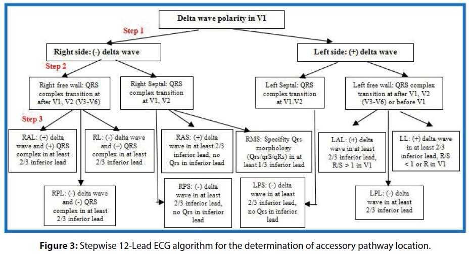
Fig. 1: Stepwise 12-Lead ECG algorithm for the determination of accessory pathway location (Source: Chu SD et al (2017).
Results
Clinical Demographics and Pathophysiological Profiles The final prospective cohort consisted of 109 patients who successfully underwent RFCA. The gender distribution was closely balanced with 56 males (51.4%) and 53 females (48.6%), showing no statistically significant difference (p > 0.05). The mean age of the population stood at 43.6 ± 14.9 years (range: 18–73 years), representing a typical adult demographic distribution. Electrophysiological and mapping diagnostics confirmed that all 109 subjects possessed a single, manifest AP. Left-sided pathways predominated slightly, accounting for 57 cases (52.3%), whereas right-sided configurations were identified in 52 cases (47.7%). The granular anatomical distribution validated by successful ablation sites is summarized in Table 1. Diagnostic Accuracy of the ECG Schematic: The pre-procedural ECG algorithm demonstrated high concordance with intraprocedural electroanatomical mapping endpoints. Overall diagnostic accuracy for correctly localizing the AP anatomical zone was 92.7% (101/109 patients). Anatomical regional accuracy was 92.3% (48/52) for right-sided APs and 93.0% (53/57) for left-sided APs.
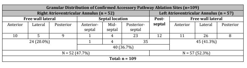
Table 1:Distribution of Accessory Pathway Locations. (A: anterior; L: lateral; P: posterior; MS: mid-septal; PS: posterior-septal)
Arrhythmia Characteristics and Procedural Safety Endpoints Table 2 shows that during the ECG study, various arrhythmias were detected, with the most common being anterograde atrial ventricular reciprocating tachycardia in 28/109 (25.7%) cases. Atrial fibrillation/flutter occurred in 3/109 (2.8%) cases. Regarding complications, no severe complications were observed, while one patient (0.9%) experienced a mild phrenic nerve response. During baseline EPS, induction protocols mapping the tachyarrhythmia profiles revealed that orthodromic atrioventricular reciprocating tachycardia (anterograde AVRT) was the primary mechanism, observed in 28 patients (25.7%). Concurrent paroxysmal atrial fibrillation or atrial flutter was induced in 3 patients (2.8%), while isolated ventricular premature beats (VPBs) occurred during catheter manipulation in another 3 cases (2.8%). The remaining 75 patients (68.8%) presented with standard manifest ventricular pre-excitation without clinical induction of sustainable sustained tachyarrhythmia circuits during the baseline EPS phase. Regarding safety endpoints, the absolute complication rate was exceptionally low. There were zero occurrences (0.0%) of major peri-procedural complications, such as cardiac tamponade, permanent complete atrioventricular block, iatrogenic valve trauma, stroke, or mortality. One transient, minor complication consisting of an intraoperative phrenic nerve response was noted in a single patient (0.9%) during ablation along the right anterior-lateral annulus; this resolved spontaneously upon immediate cessation of energy delivery without long-term sequelae. In conclusion, this study found a high prevalence of anterograde atrial ventricular reciprocating tachycardia in patients with typical WPW syndrome. The AP was predominantly located on the left side (Table 1). RFCA was effective and safe, with no severe complications.
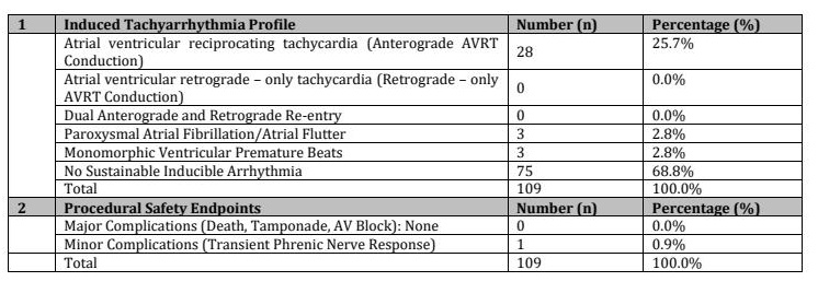
Table 2: Summary of Intraprocedural Arrhythmia Induction and Complication Profiles
Electroanatomical Mapping and Ablation Technical Dynamics The operational parameters of the RFCA procedure were sub-analyzed based on the laterality of the AP. Right-sided accessory pathways presented a significantly higher technical challenge. Total procedure time was significantly longer in the right-sided group compared to the left-sided group (52.9 ± 21.5 min vs. 44.9 ± 14.5 min, p < 0.05). Similarly, the cumulative ablation energy delivery time (368.1 ± 202.5 sec vs. 261.6 ± 110.6 sec, p < 0.01) and the total number of radiofrequency applications (10.9 ± 8.3 times vs. 5.7 ± 3.7 times, p < 0.001) were significantly elevated in the right-sided cohort. Fluoroscopic radiation exposure times were higher in right-sided interventions (7.3 ± 4.2 min) than left-sided interventions (7.0 ± 3.8 min), although this variation did not cross the threshold of statistical significance (p > 0.05).
Analysis of Safety Endpoints and Arrhythmic Complications Comparison with Historical Control Group (Table 4): To evaluate procedural trends, operational metrics of the current interventional group (n=109, managed with the predictive schematic) were compared with a historical baseline dataset (n=189, managed using conventional mapping without the schematic). The use of the ECG schematic was associated with statistically significant reductions in operational parameters. Average total procedure duration was shorter in the interventional cohort compared to the historical control group (48.7 ± 18.5 minutes vs. 55.0 ± 29.7 minutes, p < 0.05). Similarly, mean fluoroscopy exposure duration was significantly lower (7.2 ± 4.0 minutes vs. 9.0 ± 5.0 minutes, p < 0.001). Mean cumulative ablation delivery time (311.4 ± 168.2 sec vs. 314.6 ± 201.4 sec, p > 0.05) and mean number of RF applications (8.1 ± 6.7 vs. 8.3 ± 7.1, p > 0.05) trended lower in the schematic group but did not reach statistical significance. The introduction of the ECG schematic led to highly significant improvements. The average total procedure time was reduced from 55.0 ± 29.7 minutes to 48.7 ± 18.5 minutes (p < 0.05). More profoundly, the average fluoroscopy exposure duration decreased significantly from 9.0 ± 5.0 minutes down to 7.2 ± 4.0 minutes (p < 0.001). While the mean cumulative ablation delivery time (311.4 ± 168.2 sec vs. 314.6 ± 201.4 sec) and the mean number of RF applications (8.1 ± 6.7 vs. 8.3 ± 7.1) showed a downward trend in the schematic group, these specific values did not achieve standalone statistical significance (p > 0.05). The average procedure time, X-ray exposure time, ablation time, and number of ablations were significantly shorter in the group that used the prediction schematic compared to the group that did not (p < 0.05, p < 0.001, p > 0.05, and p > 0.05, respectively). This indicates that using the prediction schematic can significantly reduce procedure time, X-ray exposure, and the number of ablations, without compromising the effectiveness of the procedure.
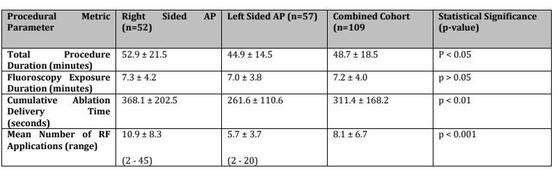
Table 3: Comparative Operational Metrics Stratified by Accessory Pathway Laterality
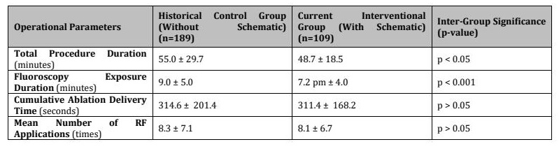
Table 4: Efficacy Analysis of the Unguided Conventional Mapping and Guided Novel Schematic Mapping
Discussion
Previous work from our group established the diagnostic performance of the ECG schematic in this prospective cohort [29]. The present analysis extends those findings by examining its potential procedural relevance. Specifically, we compared RFCA performance after implementation of the schematic with historical procedural data obtained before its introduction. This comparison suggests that schematic-guided pre-procedural localization was associated with shorter procedure and fluoroscopy times, although the historical design does not permit attribution of these differences exclusively to the algorithm
Clinical Demographics and Pathophysiological Profiles The baseline demographic architecture established in this study aligns with international cardiac electrophysiology registries, indicating that although WPW syndrome is a congenital anomaly, symptomatic exacerbation requiring definitive ablation therapy peaks during mid-adulthood [32, 33]. A key clinical finding is that older individuals within the cohort remain highly vulnerable to severe, life-threatening arrhythmias [32-35]. With advancing age, natural degenerative alterations in atrial architecture and worsening sympathetic tones increase the risk of developing rapid paroxysmal supraventricular tachycardias, dangerous atrial fibrillation, or degenerated ventricular tachycardias [32-35]. The structural distribution validated via successful ablation endpoints demonstrated a higher prevalence of left-sided APs, with a substantial concentration in the left free-wall regions, which is consistent with the established literature [36-39]. Regarding arrhythmia presentation during diagnostic pacing, the high prevalence of orthodromic AVRT (25.7%) highlights its status as the classic clinical manifestation of macro-reentrant pathways [37, 38]. Importantly, our findings regarding induced atrial fibrillation/flutter (2.8%) underscore the necessity of early therapeutic interventions [32, 34]. In patients demonstrating rapid anterograde conduction over the AP, AF carries an immediate risk of precipitating hemodynamic collapse or degenerate ventricular fibrillation [11, 32, 34]. In our study, patients who experienced transient episodes of AF or rapid tachycardia during catheter manipulation were safely converted back to stable sinus rhythm using synchronized electrical cardioversion or immediate localized cooling. Post-ablation pacing confirmed long-term electrical stabilization with no recurrence of tachyarrhythmias.
Electroanatomical Mapping and Ablation Technical Dynamics Endocardial localization of the AP involves precise coordination between non-invasive ECG predictions and invasive catheter feedback [14-16, 31]. The standard mapping approach utilizes multipolar catheters introduced via the femoral veins to map the tricuspid valve circumference for right-sided pathways [31]. For left-sided free wall or lateral pathways, a retrograde transaortic approach via the femoral artery into the left ventricle is typically preferred, allowing the catheter tip to be stabilized beneath the mitral valve leaflets [31, 44, 47]. Ablation is directed at target sites demonstrating the maximum advancement of local activation signals relative to surface electrical vectors [31, 40]. The total operational time required to achieve successful ablation is multi-factorial, depending heavily on the anatomical site of the pathway, the experience of the interventional team, and the availability of localization tools [40-41]. Early historical cohorts reported prolonged procedure and radiation times. For instance, landmark trials by Lemery et al. (1992) and Calkins et al. (1992) documented mean procedure times of 216.0 ± 90.0 minutes and 134.0 ± 75.0 minutes, respectively, with corresponding fluoroscopy times reaching 66.0 ± 33.0 and 47.0 ± 33.0 minutes [42, 43]. Over time, technological improvements and increased operator experience have steadily reduced these parameters, as shown by Schwagten et al. (2010), who reported an optimized mean procedure duration of 87.1 ± 30.8 minutes and a fluoroscopy time of 14.4 ± 4.7 minutes [44]. In historical studies from Canada, such as Dubuc et al., early mapping strategies achieved success rates of 93.3% but required significantly longer procedural sessions [45]. On Diagnostic Accuracy & Procedural Efficiency. In comparison, the current prospective study achieved a highly optimized mean total procedure time of 48.7 ± 18.5 minutes and a mean fluoroscopy exposure time of 7.2 ± 4.0 minutes. In the present study, the prospective cohort achieved a mean total procedure time of 48.7 ± 18.5 minutes and a mean fluoroscopy exposure time of 7.2 ± 4.0 minutes. The high diagnostic accuracy of the novel ECG schematic (92.7% concordance with mapping endpoints) likely facilitated these observed improvements [27, 28, 40, 46]. By identifying the target anatomical quadrant prior to vascular access, the algorithm aids in optimizing catheter selection and initial positioning, thereby reducing unnecessary endocardial mapping and curtailing ionizing radiation exposure for both patients and medical staff [27, 28, 41, 47]. From a radiation safety perspective, minimizing fluoroscopy time is critical. Chronic radiation exposure is linked to cumulative chromosomal damage, skin injuries, and increased malignancy risks for interventional specialists [20]. Our achieved mean exposure time of 7.2 minutes falls well within established international safety guidelines, demonstrating excellent radioprotective efficacy [20]. Our sub-analysis based on pathway laterality revealed that right-sided APs required significantly longer procedure durations (52.9 ± 21.5 min and 44.9 ± 14.5 min, p < 0.05) and increased radiofrequency energy applications (10.9 ± 8.3 and 5.7 ± 3.7, p < 0.001) compared to left-sided pathways. This disparity is driven by anatomical and mechanical factors [31]. The right atrium and tricuspid annulus lack the rigid structural support found in the left ventricle, making catheter stabilization difficult due to continuous cardiac and respiratory movements [31]. Additionally, right-sided pathways often exhibit greater structural complexity and broader fibers, requiring multiple consolidation lesions to achieve permanent conduction blocks [31]. By providing highly accurate lateralization, the novel ECG schematic helps operators anticipate these challenges, select appropriate vascular access routes, and optimize initial catheter positioning [27, 28, 41, 47]. In many cases, this precise localization enabled immediate pathway elimination within the first 1 to 2 seconds of radiofrequency energy delivery [41, 47].
Analysis of Safety Endpoints and Arrhythmic Complications During baseline EPS, induction protocols mapping the tachyarrhythmia profiles revealed that orthodromic atrioventricular reciprocating tachycardia (anterograde AVRT) was the primary mechanism, observed in 28 patients (25.7%). Concurrent paroxysmal atrial fibrillation or atrial flutter was induced in 3 patients (2.8%), while isolated ventricular premature beats (VPBs) occurred during catheter manipulation in another 3 cases (2.8%). The remaining 75 patients (68.8%) presented with standard manifest ventricular pre-excitation without clinical induction of sustainable sustained tachyarrhythmia circuits during the baseline EPS phase. Regarding safety endpoints, the absolute complication rate was exceptionally low. There were zero occurrences (0.0%) of major peri-procedural complications, such as cardiac tamponade, permanent complete atrioventricular block, iatrogenic valve trauma, stroke, or mortality. One transient, minor complication consisting of an intraoperative phrenic nerve response was noted in a single patient (0.9%) during ablation along the right anterior-lateral annulus; this resolved spontaneously upon immediate cessation of energy delivery without long-term sequelae. In conclusion, this study found a high prevalence of anterograde atrial ventricular reciprocating tachycardia in patients with typical WPW syndrome. The AP was predominantly located on the left side (Table 1). RFCA was effective and safe, with no severe complications. A critical metric of any novel diagnostic algorithm is its capacity to enhance procedural safety [20]. In our cohort, the absence of major complications (0.0%), including structural atrioventricular blocks, stands in favorable contrast to historical literature [48]. Prior multicenter registries, such as the Pediatric Radiofrequency Ablation Registry reported by Schaffer et al. (1996), documented high rates of inadvertent AV block, particularly when treating pathways near sensitive conduction tissue (10.4% in mid-septal and 2.7% in anterior-septal locations) [48]. Mandapati et al. (2003) similarly noted permanent AV block rates of up to 10% in mid-septal interventions. The zero-incidence rate of complete heart block in our study, even for pathways close to the His bundle or AV node, reflects the high spatial precision provided by the ECG schematic [41]. By mapping the boundaries of the septal matrix accurately, the algorithm allowed operators to apply highly targeted energy delivery, avoiding structural damage to the normal conduction pathway [41]. The single case of transient phrenic nerve response (0.9%) resolved immediately upon turning off the RF generator, confirming that the procedural protocol maintains an exceptional safety margin. The prediction schematic significantly reduced procedure time, X-ray exposure time, and the number of ablations (Table 3). This is because the schematic helped guide the decision on whether to approach the AP through the femoral artery or vein and also provided detailed information about the AP's location. This allowed for more efficient mapping and ablation, reducing procedure time and radiation exposure [27, 28, 41, 47]. While the prediction schematic showed significant improvements, further research is needed to determine if other factors, such as the experience of the interventional cardiologist, the type of equipment used, and the specific characteristics of the AP, could also influence procedure time and outcomes. When applying the prediction schematic for AP location (Schematic 1), accurately predicting the AP on the right or left side can guide the decision on whether to insert the mapping electrode through the femoral vein or artery [27, 28, 41, 47]. The detailed prediction of the AP's location within the septal region (anterior, mid-septal, or posterior) can help quickly guide the electrode to the target area within 1-2 cm of the valve [41]. This can anticipate potential challenges during the procedure and reduce the risk of complications associated with ablating APs in the anterior septal and mid-septal regions, especially those located near the His bundle and atrioventricular node [41]. The prediction schematic allowed us to quickly reach the target ablation site, and in many cases, the AP was successfully ablated within the first 1-2 seconds (Fig. 2) [47].
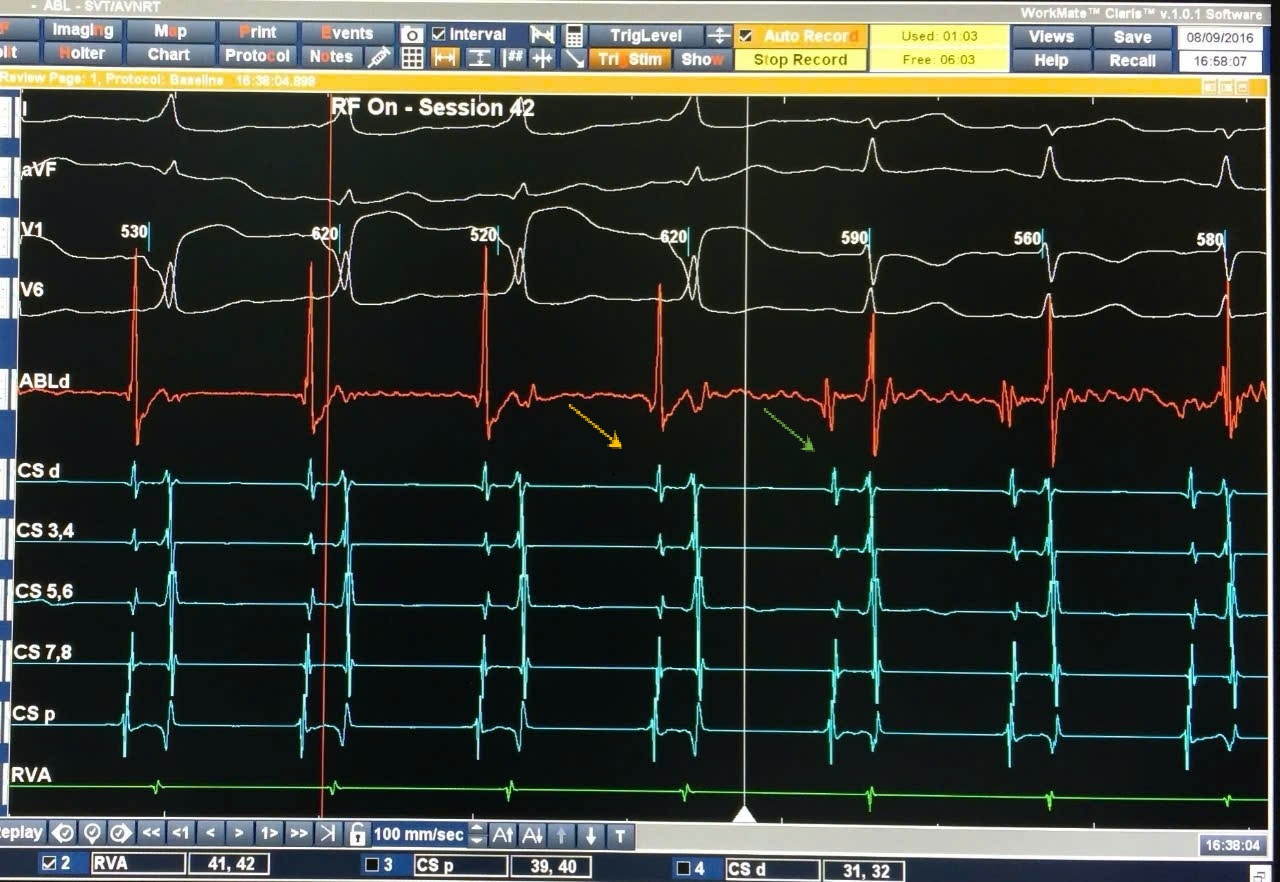
Fig. 2: ECG before ablation with target point (Yellow arrow) and successful RFCA of Accessory pathway in the first second time (Blue arrow). (Source: A female patient-27 years with right accessory pathway, Chu SD et al (2018) [47].
Methodological Considerations and Study Limitations Several methodological aspects require cautious interpretation. First, because the comparison relies on a historical control cohort (n=189), direct causality between the implementation of the ECG schematic and improved procedural parameters cannot be definitively established. While the observed reductions in procedure and fluoroscopy times were statistically significant, progressive operator experience, subtle procedural refinements, and incremental advances in catheter manipulation techniques over time may also have contributed to these improved operational metrics. Furthermore, this investigation was conducted at a single tertiary center with a sample size of 109 patients in the prospective arm, which limits our ability to evaluate the algorithm's performance across rarer or more complex anatomical AP sub-types. Additionally, the study did not include a concurrent randomized control group, relying instead on historical control data for efficacy comparisons. Future prospective, randomized controlled trials comparing schematic-guided mapping directly against conventional mapping strategies are warranted to confirm these findings and isolate the independent effect of the diagnostic algorithm.
Conclusion
This prospective study indicates that the novel 12-lead surface ECG prediction schematic offers accurate pre-procedural localization of accessory pathways in typical Wolff–Parkinson–White syndrome, achieving an overall diagnostic accuracy of 92.7%. Its clinical deployment was associated with significant reductions in total procedure duration and fluoroscopy exposure times while maintaining a 100% acute procedural success rate and a favorable safety profile with no major complications. Given these findings, integrating this structured ECG schematic into routine pre-procedural planning may serve as a useful adjunctive tool to streamline electrophysiological mapping in patients with typical WPW syndrome.
Acknowledgements
Author Contributions
All authors contributed equally to the study design, methodology, execution, data analysis, manuscript drafting, and editing.
Disclosure Statement
The authors have nothing to disclose.
References
- Papadakis MA, McPhee SJ, Rabow MW (2016). Current Medical Diagnosis and Treatment 2016, Lange, McGraw-Hill Education.
- Surawicz B, Knilans TK, Te-Chuan C (2008). Chou's Electrocardiography in Clinical Practice: Adult and Pediatric, Elsevier Saunders, Philadelphia, PA.
- Dong VT (2006). Treatment of Wolff-Parkinson-White Syndrome with Radiofrequency Energy. Dissertation, Vietnam Military Medical Academy.
- Narula OS (1973). Wolff-Parkinson-White Syndrome: A review. Circulation, 47, 872-887. https://doi.org/10.1161/01.CIR.47.4.872
- Olshausen KV, Le MK, Nguyen SH (2016). ECG from Basic to Advanced. Medical Publishing House, Ho Chi Minh City Branch.
- Schaider JJ, Barkin RM, Hayden SR, et al. (2015). Rosen & Barkin's 5-Minute Emergency Medicine Consult, Wolters Kluwer Health/Lippincott Williams & Wilkins, Philadelphia.
- Kulig J, Koplan BA (2010). Cardiology patient page: Wolff-Parkinson-White syndrome and accessory pathways. Circulation, 122, e480-e483. https://doi.org/10.1161/CIRCULATIONAHA.109.929372
- Cahyaningtias M, Hanafy DA (2011). Wolff-Parkinson-White syndrome in young men presenting with palpitation: The pattern of delta waves in predicting location of accessory pathway. Med J Indones, 20, 298-301. https://doi.org/10.13181/mji.v20i4.462
- Cobb FR, Blumenschein SD, Sealy WC, et al. (1968). Successful surgical interruption of the bundle of Kent in a patient with Wolff-Parkinson-White syndrome. Circulation, 38(6), 1018-1029. https://doi.org/10.1161/01.CIR.38.6.1018
- Hanon S, Shapiro M, Schweitzer P (2005). Early history of the pre-excitation syndrome. Europace, 7, 28-33. https://doi.org/10.1016/j.eupc.2004.09.005
- Obeyesekere M, Gula LJ, Skanes AC, et al. (2012). Risk of sudden death in Wolff-Parkinson-White syndrome: How high is the risk? Circulation, 125(5), 659-660. https://doi.org/10.1161/CIRCULATIONAHA.111.085159
- Thakur RK, Klein GJ, Yee R (1994). Radiofrequency catheter ablation in patients with Wolff-Parkinson-White syndrome. CMAJ, 151(6), 771-776.
- Sundaram PS, Sra J (2015). Utility of unipolar recordings for complex Wolff-Parkinson-White ablation. Indian Pacing Electrophysiol J, 15(2), 125-129. https://doi.org/10.1016/j.ipej.2015.07.010
- Milstein S, Sharma AD, Guiraudon GM, et al. (1987). An algorithm for the electrocardiographic localization of accessory pathways in the Wolff-Parkinson-White syndrome. Pacing Clin Electrophysiol, 10(1), 555-563. https://doi.org/10.1111/j.1540-8159.1987.tb04520.x
- Fitzpatrick AP, Gonzales RP, Lesh MD, et al. (1994). New algorithm for the localization of accessory atrioventricular connections using a baseline electrocardiogram. J Am Coll Cardiol, 23(1), 107-116. https://doi.org/10.1016/0735-1097(94)90508-8
- Arruda MS, McClelland JH, Wang X, et al. (1998). Development and validation of an ECG algorithm for identifying accessory pathway ablation site in Wolff-Parkinson-White syndrome. J Cardiovasc Electrophysiol, 9(1), 2-12. https://doi.org/10.1111/j.1540-8167.1998.tb00861.x
- Eisenberger M, Davidson NC, Todd DM, et al. (2010). A new approach to confirming or excluding ventricular preexcitation on a 12-lead ECG. EP Europace, 12(1), 119-123. https://doi.org/10.1093/europace/eup345
- Taguchi N, Yoshida N, Inden Y, et al. (2014). A simple algorithm for localizing accessory pathways in patients with Wolff-Parkinson-White syndrome using only the R/S ratio. Journal of Arrhythmia, 30(6), 484-489. https://doi.org/10.1016/j.joa.2013.10.006
- Singer I, Kupersmith J (1993). Clinical Manual of Electrophysiology, Williams & Wilkins, Baltimore.
- Cohen MI, Triedman JK, Cannon BC, et al. (2012). PACES/HRS expert consensus statement on the management of the asymptomatic young patient with a Wolff-Parkinson-White electrocardiographic pattern. Heart Rhythm, 9(6), 1006-1024. https://doi.org/10.1016/j.hrthm.2012.03.050
- Miklos SS, Szliagyi L, Gorog LK, et al. (2011). An enhanced method for accessory pathway localization in case of Wolff-Parkinson-White syndrome. Acta Physiol Hung, 98(3), 347-358. https://doi.org/10.1556/APhysiol.98.2011.3.12
- Akobeng AK (2007). Understanding diagnostic tests 1: Sensitivity, specificity and predictive values. Acta Paediatrica, 96, 338-341. https://doi.org/10.1111/j.1651-2227.2006.00180.x
- Parikh R (2008). Understanding diagnostic tests 1: Sensitivity, specificity and predictive values. Indian Journal of Ophthalmology, 56(1), 45-50. https://doi.org/10.4103/0301-4738.37595
- Tilaki KH (2014). Sample size estimation in diagnostic test studies of biomedical informatics. Journal of Biomedical Informatics, 48, 193-204. https://doi.org/10.1016/j.jbi.2014.02.013
- Bujang MA, Adnan TH (2016). Requirements for minimum sample size for sensitivity and specificity analysis. J Clin Diagn Res, 10(10), YE01-YE06. https://doi.org/10.7860/JCDR/2016/18129.8744
- Zaidi SMH, Waseem HF, Ansari FA, et al. (2016). Sample size estimation of diagnostic test studies in health sciences. Proc. 14th International Conference on Statistical Sciences, 29, 239-246.
- Si CD, Khanh PQ, Dong TV (2017). 12 Lead electrocardiogram features of accessory pathways localization in typical Wolff-Parkinson-White syndrome patients - 3rd Global Summit on Heart Diseases 2017 J Clin Exp Cardiolog, 8:10 (Suppl).
- Chu SD, Pham KQ, Tran DV (2017). 12 Lead electrocardiogram algorithm for the localization of accessory pathways using simple parameters in patients with typical Wolff-Parkinson-White syndrome. Interventional Cardiology, 9 (6), 229-237. https://doi.org/10.4172/Interventional-Cardiology.1000585
- Chu SD, Pham KQ, Tran DV (2017). Accuracy of the new electrocardiogram algorithm in predicting localization of accessory pathways in patients with typical Wolff - Parkinson - White syndrome. Interventional Cardiology, 9 (6), 243-249 https://doi.org/10.4172/Interventional-Cardiology.1000587
- Dar MA, Sheikh SH, Abid AR, et al. (2008). Localization of accessory pathways according to AP Fitzpatrick ECG criteria in patients with Wolff-Parkinson-White syndrome in our population. Pakistan Heart Journal, 41(3-4), 22-27.
- Murgatroyd FD, Krahn AD, Klein GJ, et al. (2008). Handbook of Cardiac Electrophysiology: A Practical Guide to Invasive EP Studies and Catheter Ablation, Remedica Publishing Limited, London.
- Brembilla-Perrot B (2008). Management of Wolff-Parkinson-White syndrome in the elderly. European Cardiology, 4, 72-74. https://doi.org/10.15420/ecr.2008.4.2.72
- Brembilla-Perrot B, Holban I, Houriez P, et al. (2001). Influence of age on the potential risk of sudden death in asymptomatic Wolff-Parkinson-White syndrome. Pacing Clin Electrophysiol, 24, 1514-1518. https://doi.org/10.1046/j.1460-9592.2001.01514.x
- Dagres N, Hindricks G, Kottkamp H, et al. (2001). Impact of radiofrequency catheter ablation of accessory pathways on the frequency of atrial fibrillation during long-term follow-up: High recurrence rate of atrial fibrillation in patients older than 50 years of age. Eur Heart J, 22, 423-427. https://doi.org/10.1053/euhj.2000.2429
- Karavidas A, Lazaros G, Tsiachris D, et al. (2010). Aging and the cardiovascular system. Hellenic Journal of Cardiology, 51, 421-427.
- Liebman J, Zeno JA, Olshansky B, et al. (1991). Electrocardiographic body surface potential mapping in the Wolff-Parkinson-White syndrome: Noninvasive determination of the ventricular insertion sites of accessory atrioventricular connections. Circulation, 83(3), 886-901. https://doi.org/10.1161/01.CIR.83.3.886
- Al-Zaiti SS, Magdic KS (2016). Paroxysmal supraventricular tachycardia: Pathophysiology, diagnosis, and management. Crit Care Nurs Clin North Am, 28(3), 309-316. https://doi.org/10.1016/j.cnc.2016.04.005
- Sethi KK, Dhall A, Chadha DS, et al. (2007). WPW and preexcitation syndromes. J Assoc Physicians India, 55 Suppl, 10-15.
- Teixeira CM, Pereira TA, Lebreiro A, et al. (2016). Accuracy of the electrocardiogram in localizing the accessory pathway in patients with Wolff-Parkinson-White pattern. Arq Bras Cardiol, 107(4), 331-338. https://doi.org/10.5935/abc.20160132
- Morady F (1999). Drug therapy: Radio-frequency ablation as treatment for cardiac arrhythmias. The New England Journal of Medicine, 340(7), 534-544 .https://doi.org/10.1056/NEJM199902183400707
- Chu SD, Pham KQ, Tran DV (2018). Development and evaluation of surface electrocardiogram in the septal accessory pathway localization in typical Wolff-Parkinson-White syndrome. Biomedical Research and Therapy, 5(9), 2680-2687. https://doi.org/10.15419/bmrat.v5i9.479
- Lemery R, Talajic M, Roy D, et al. (1992). Success, safety, and late electrophysiological outcome of low-energy direct-current ablation in patients with the Wolff-Parkinson-White syndrome. Circulation, 85(3), 957-962. https://doi.org/10.1161/01.CIR.85.3.957
- Calkins H, Langberg J, Sousa J, et al. (1992). Radiofrequency catheter ablation of accessory atrioventricular connections in 250 patients: Abbreviated therapeutic approach to Wolff-Parkinson-White syndrome. Circulation, 85(4), 1337-1346. https://doi.org/10.1161/01.CIR.85.4.1337
- Schwagten B, Jordaens L, Ayerza MR, et al. (2010). A randomized comparison of transseptal and transaortic approaches for magnetically guided ablation of left-sided accessory pathways. Pacing Clin Electrophysiol, 33, 1298-1303. https://doi.org/10.1111/j.1540-8159.2010.02810.x
- Dubuc M, Nadeau R, Tremblay G, et al. (1993). Pace mapping using body surface potential maps to guide catheter ablation of accessory pathways in patients with Wolff-Parkinson-White syndrome. Circulation, 87(1), 135-143. https://doi.org/10.1161/01.CIR.87.1.135
- Igor S (2001). Electrophysiologic study. In Igor Singer: Clinical Manual of Electrophysiology, Lippincott Williams & Wilkins, Philadelphia, USA, 52-68.
- Schaffer MS, Silka MJ, Ross BA, et al. (1996). Inadvertent atrioventricular block during radiofrequency catheter ablation. Results of the Pediatric Radiofrequency Ablation Registry. Circulation, 94(12), 32 https://doi.org/10.1161/01.CIR.94.12.3214
- Chu SD, Pham KQ, Tran DV (2018). Development and evaluation of 12-lead electrocardiogram in the left free wall of accessory pathway localization in patients with typical Wolff-Parkinson-White syndrome. Biomedical Research and Therapy, 5(11), 2832-2840. https://doi.org/10.15419/bmrat.v5i11.502