Corresponding Author: Yongqiu Zheng

Inst. of Basic Med. Sciences of Xiyuan Hospital China Academy of Chinese Med. Sciences, Beijing 100091 (China)
E-Mail yongqiuzheng@sina.com


Erratum

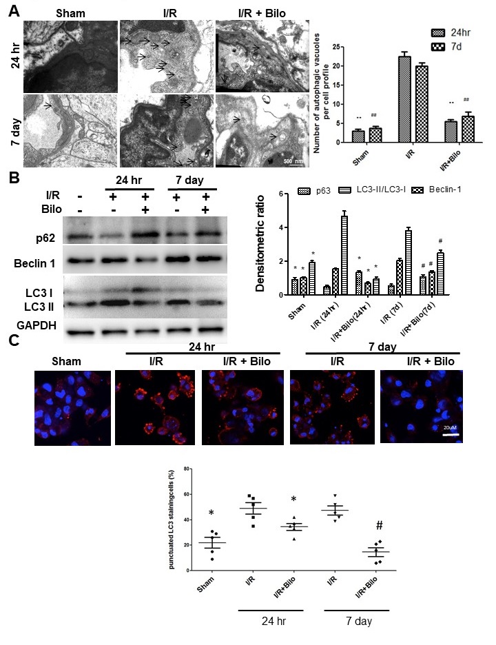
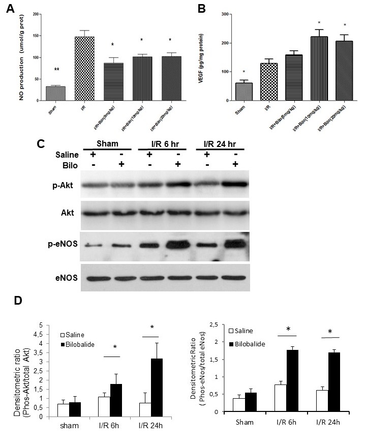
In the article by Zheng, et al., entitled “By Activating Akt/eNOS Bilobalide B Inhibits Autophagy and Promotes Angiogenesis Following Focal Cerebral Ischemia Reperfusion” [Cell Physiol Biochem 2018;47:604-616, DOI: 10.1159/000490016], due to carelessness during manuscript preparation, wrong images have been used for Fig. 4C (I/R+Bilo 24 hr and I/R 7 day) and Fig. 5C (p-eNOS and total eNOS) and would like to correct this. Although some data for Fig. 4C have been lost, the authors have been able to re-perform the staining of LC3 with newly cutted slides from the original tissue blocks and therefore have changed all images in Fig. 4C for consistency.
The authors confirm that the updated data is similar to their original data and still support the results and conclusions of their paper. The corrected Fig. 4 and Fig. 5 are displayed below.

Fig. 4. Bilobalide treatment suppresses autophagy following I/R injury to the brain. (A) Representative electron microscope images show that I/R injury induced formation of autophagic vacuoles (identified by arrows), but that treatment with bilobalide (10mg/ kg) could inhibit their formation. Summary of upper panels was showed in lower panel. (B) Western blot analysis showed that treatment with bilobalide (10mg/ kg) significantly suppressed Beclin-1 and the LC3-II/LC3-I ratio, two key autophagy related markers, following I/R injury. (C) LC3 foci formation (foci showed in red) was visualized using a Zeiss LSM- 510 microscope (upper panels) and quantified by Image-Pro Plus (lower panel). Treatment with bilobalide (10mg/kg) reduced LC3 foci formation induced by I/R injury. Data are presented as mean±SD. n=12 rats per group at 24 hour time point; n=10 rats per group at 7 day time point. ΔΔ, ## or ** : p<0.01; Δ, # or * : p<0.05.

Fig. 5. Bilobalide treatment induces NO and VEGF production through activation of eNOS and Akt pathways. (A) Nitric oxide (NO) and (B) VEGF production from infarcted areas of the brain were measured by ELISA analysis. Bilobalide treatment inhibited NO and promoted VEGF production. (C) Western blot analysis showed that bilobalide (10mg/kg) treatment enhanced p-Akt and p-eNOS levels. Results were obtained from three independent experiments; only one representative experiment is shown. (D) Quantitative densitometry of western blots presented in (C). Data are presented as mean±SD. n=12 rats per group at 24 hour time point; n=10 rats per group at 7 day time point. ** : p<0.01; *: p<0.05.