Corresponding Author: Yumin Li

The Second Hospital of Lanzhou University, Lanzhou, Gansu Province, 730030 (China)
E-Mail liym@lzu.edu.cn


Erratum

The authors of the original article by Qi, et al., entitled “Exosomes Derived from Human Bone Marrow Mesenchymal Stem Cells Promote Tumor Growth Through Hedgehog Signaling Pathway” [Cell Physiol Biochem 2017;42(6):2242-2254, DOI: 10.1159/000479998], would like to address two issues which have been raised on their platform PubPeer against two figures, Fig. 2 and Fig. 4B, in their paper.


For the issue about Fig. 2, the authors would like to respond as follows:
“Identification of exosomes by morphology (e.g., using EM) or by examining the exosome markers using WB can be difficult and sometimes unreliable. In our study, EM immunostaining was not done due to the lack of facility. However, our WB data for the exosome marker CD63 did show some “smearing”, albeit faint in the Figure. We believe this was likely due to low concentration of the target protein (CD63) and low resolution of the WB photo.”

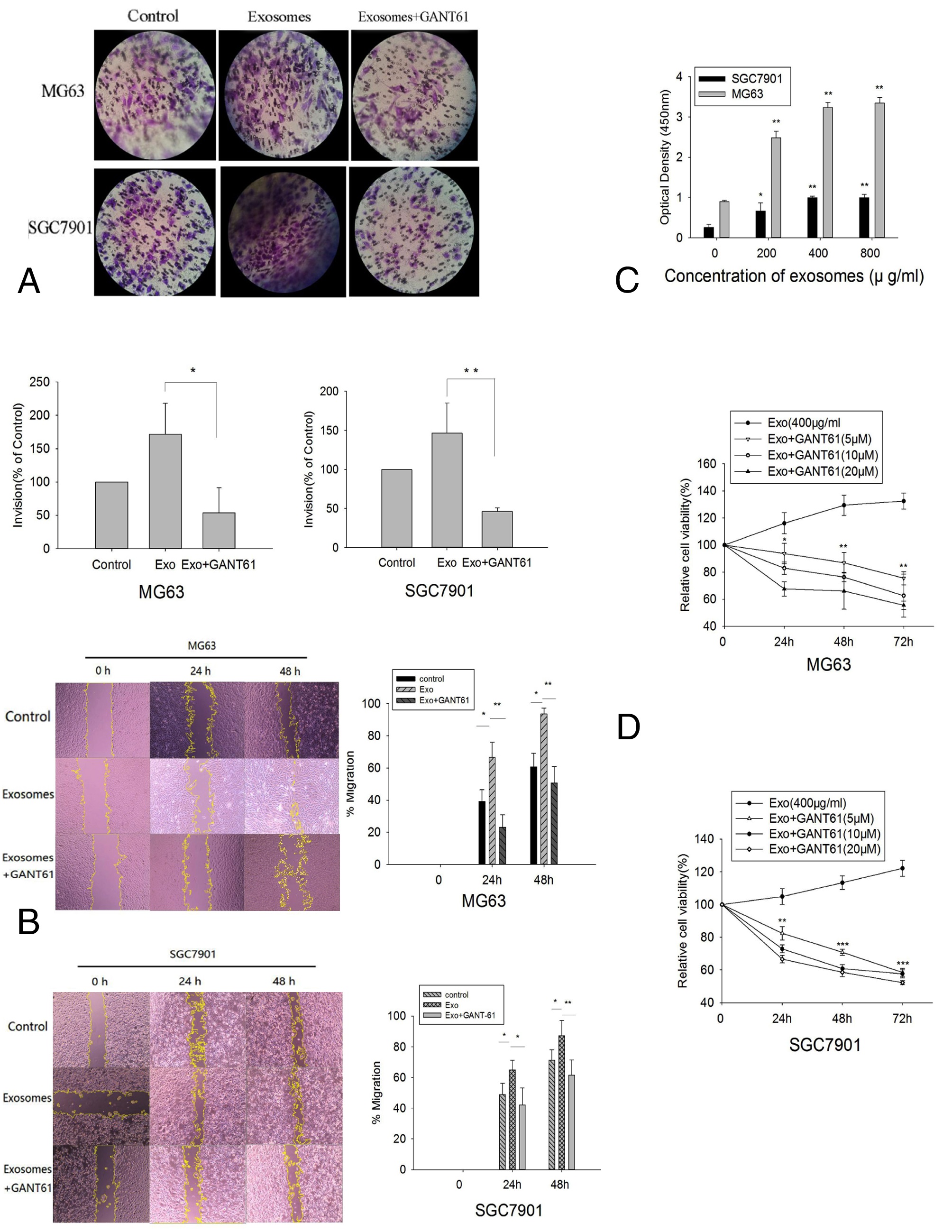
For the issue about Fig. 4, they would like to display the correct Fig. 4 (see next page), because they have identified that two images in Fig. 4B were mistakenly used. They also confirm that the image for Exosome 0h (SGC7901) ist correct, although it is turned 90 degrees.

The authors confirm that all of the results and conclusions of the article remain unchanged, as well as the figure legend.

Fig. 4. hBMSC-exosomes induce viability and proliferation in MG63 and SGC7901 cells. (A) an amounts of MG63 cells and SGC7901 cells were respectively added to the upper chamber of transwell with matrigel coated membrane. Cancer cells were treated with exosomes (400µg/ml) or treated with exosomes (400µg/ ml) and GANT-61(10µM), an equal volume of exosome-depleted medium was used as a control. After 24 hours the number of cells migrated to the lower chamber of the 8 µm pore-sized membrane were analyzed by taking photos and counting the number of cells per visual field. n=3 per group; *P <0.05, **P <0.01. (B) Scratch migration assay test of interfering hBMSC-exosomes group, interfering GANT61 group and control group in 24 hours and 48 hours. The wound healing assay demonstrated a stronger migration ability of cells in interfering hBMSC-exosomes group. Compared with control group and interfering GANT61 group, there were significant difference in percentage of wound closed(n=3 per group; *P <0.05, **P <0.01).a weaker migration ability of cells in the blank control and GANT61 group. (C) MG63 and SGC7901 cells were respectively co-cultured with different concentrations of hBMSC-exosomes (0, 200, 400 and 800µg/ml) for 24 hours and then subjected to CCK-8 analyses. n=3 per group; *P <0.05, **P <0.01. (D) MG63 or SGC7901 cells in serum-free medium were treated with 400µg/ml hBMSC-exosomes or hBMSC-exosomes and different concentrations of GANT-61(0, 5, 10, 20 µM) in a 96-well plates. Cell viability was measured using CCk-8 analyses at 24, 48 and 72 hours after exosomes and GANT-61 treatment. n=3 per group; *P <0.05, **P <0.01, ***P<0.001.