Corresponding Author: Yong Zhang and Baofeng Yang

Department of Pharmacology, College of Pharmacy, Harbin Medical University, Baojian Road 157, Harbin, Heilongjiang (China)
Tel. +86 451 86671354, E-Mail hmuzhangyong@hotmail.com; yangbf@ems.hrbmu.edu.cn


Erratum

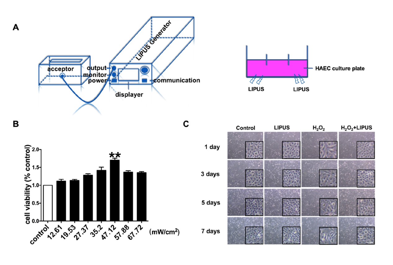
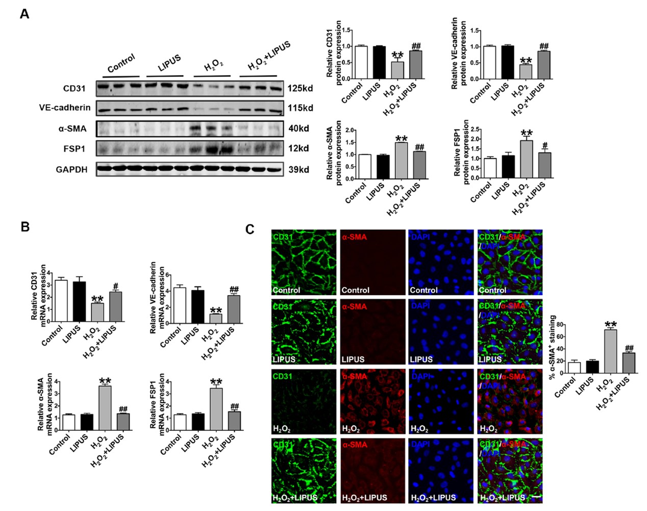
In the article “Low-Intensity Pulsed Ultrasound Prevents the Oxidative Stress Induced Endothelial- Mesenchymal Transition in Human Aortic Endothelial Cells” [Cell Physiol Biochem 2018;45:1350-1365, DOI: 10.1159/000487561] by Li et al., the incorrect representative images were mistakenly included for Figure 1C 3 days H2O2+LIPUS and Figure 2C H2O2. The corrected Figure 1 and Figure 2 are shown here.

Fig. 1. Effect of LIPUS on viability of human aortic endothelial cells (HAECs). (A) Schematic illustration of the experimental prodcedures and the customized ultrasound stimulation system. (B) Cultured HAECs were exposed to different intensities of LIPUS (12.61-67.72 mW/cm2) for seven days. Cell viability as measured by MTT assay. **p<0.01 vs Control, n =4, ANOVA, mean ± SEM. (C) LIPUS was applied to the HAECs at an intensity of 47.12mW/cm2 for 24 hours after the beginning of cell culture and then the second to the seventh LIPUS sessions were carried out every 24 h, n =4, scale bar: 50.0 μm and 200.0 μm.
Fig. 2. The effects of LIPUS on EndMT in HAECs. (A-B) Effects of LIPUS on the expression of CD31, VE-cadherin, α-SMA and FSP1 at mRNA and protein levels in the four groups: control, LIPUS, H2O2, H2O2+LIPUS. **p<0.01 vs control; #p <0.05, ##p<0.01 vs H2O2 (100 μm), n = 4-6, mean ± SEM. GADPH was used as an internal control. (C) Immunofluorescence results indicating the localization of endothelial markers CD31 (green) and mesenchymal markers α-SMA (red) in the normal group (control), treated with H2O2 (100 μm) for seven days and co-treated with LIPUS (ultrasound at 47.12 mW/cm2) and H2O2 (100 μm) for seven days. Nuclear staining DAPI (Blue), scale bar: 50 μm. **p<0.01 vs control; ##p<0.01 vs H2O2.