×
![]()
Corresponding Author: Li Zheng
The Medical and Scientific Reserch Center, Guangxi Medical University, Nanning 530021 (China)
E-Mail zhengli224@163.com
Erratum
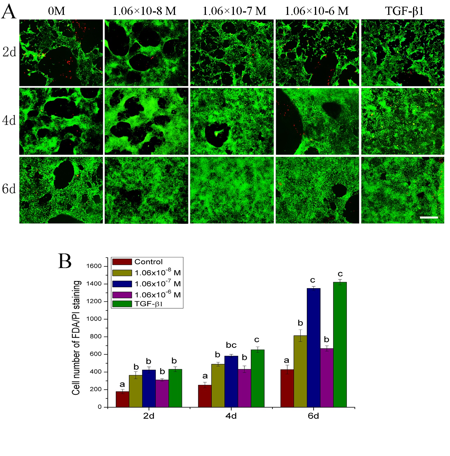
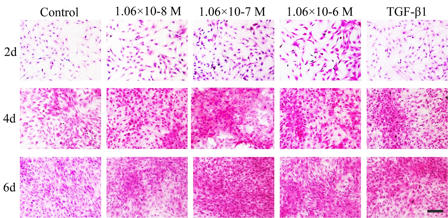
In the article “Stimulating Effect of a Newly Synthesized Sulfonamido-Basedgallate on Articular
Chondrocytes in Vitro” [Cell Physiol Biochem 2015;37:1196-1209; DOI: 10.1159/000430243]
by Lu et al., incorrect representative images were included in Figure 3A (1.06x10⁻⁸ M at day 2,
0 M at day 4, 1.06x10⁻⁸ M at day 4, 1.06x10⁻⁷ M at day 4 and 1.06x10⁻⁸ M at day 6) and Figure 4
(1.06x10⁻⁷ M at day 4). This was as a result of poor record keeping at the time of the experiment.
The correct files have been verified by review of the laboratory notebooks and the authors state
that the results and conclusions of the article remain unchanged. The authors apologize for any
inconvenience caused.
The corrected Figure 3 and Figure 4 are shown here. The corrected Figure 3 includes images
corrected in a previous Erratum (https://doi.org/10.33594/000000137).