×
![]()
Corresponding Author: Anjaneyulu Kowluru
Biomedical Research Service, B4237, John D. Dingell VA Medical Center, 4646 John R, Detroit, MI 48201 (USA)
Tel. +1-313-576-4478, Fax +1-313-576-1112 , E-Mail akowluru@med.wayne.edu
Hyperglycemic Conditions Promote Rac1-Mediated Serine536 Phosphorylation of p65 Subunit of NFκB (RelA) in Pancreatic Beta Cells
Anjaneyulu Kowlurua,b Suhadinie Gamagea,b Mirabela Halia,b Noah Gleasona,b
aBiomedical Research Service, John D. Dingell VA Medical Center, Detroit, MI, USA, bDepartment of Pharmaceutical Sciences, Eugene Applebaum College of Pharmacy and Health Sciences, Wayne State University, Detroit, MI, USA
Introduction
It is well established that exposure of pancreatic islet beta cells to metabolic stress conditions (e.g., high glucose, saturated fatty acids, biologically active sphingolipids, and pro-inflammatory cytokines) results in significant alterations in cellular function, including induction of oxidative and endoplasmic reticulum (ER) stress, stress kinase activation, mitochondrial dysfunction, and nuclear collapse leading to cell demise [1-10]. Several underlying signaling pathways have been proposed, including induction of apoptotic genes, in the cascade of events leading to dysfunction of the islet beta cells under metabolic stress [11-16]. Along these lines existing evidence supports key roles for NFκB, a transcription factor, in the regulation of cellular function under conditions of stress, inflammation and pathology of various diseases [17-23]. Published evidence also implicates NFκB in regulation of islet beta cell function in health and diabetes [19, 24-26].
NFκB is localized, in its inactive state, in the cytosolic compartment as a p65/p50 heterodimer via complexation with IκB proteins. Under conditions of increased intracellular stress and inflammation, NFκB gains its active conformation following a signaling step involving phosphorylation of IκB, which, in turn, releases p65 (encoded by the RelA gene) leading to translocation of NFκB to the nuclear compartment for induction of specific genes involved in stress/inflammation-mediated cellular dysregulation and demise [18, 20, 27]. Besides IκB, the p65 subunit of NFκB is functionally regulated via phosphorylation at its critical S276 and S536 residues. Evidence in multiple cell types suggests that phosphorylation of p65 at Ser276 is mediated by protein kinase A and ribosomal protein S6 kinase alpha-5 (MSK1) kinase in the cytosolic and nuclear compartments, respectively. IκB kinase (IKK), TANK-binding kinase 1 (TBK1), and 90 kDa ribosomal S6 kinase (RSK1) have been identified as putative kinases that control the phosphorylation of p65 at the S536 residue [27-32]. Lastly, it is widely felt that phosphorylation at S276 promotes half-life of p65, while activation of S536 results in increased proteasomal degradation of NFκB; based on these conclusions, it is postulated that phosphorylation of p65 at S276 contributes to cell survival, whereas phosphorylation at S536 accelerates cell death via apoptosis [33].
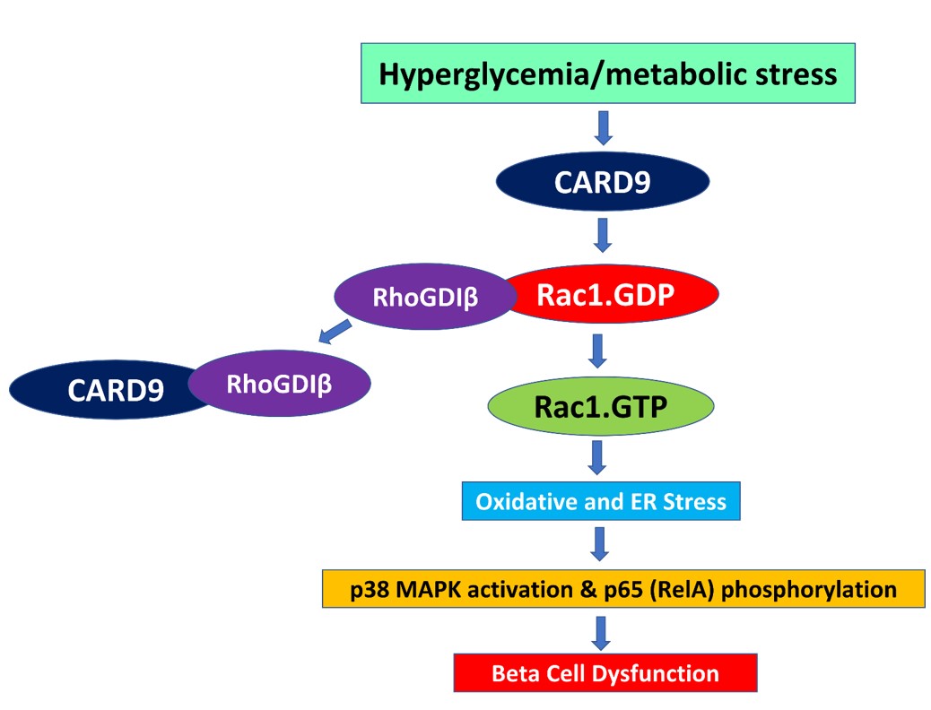
Recent investigations from our laboratory revealed that caspase recruitment domain family member 9 (CARD9) mediates metabolic dysfunction of the pancreatic beta cell via activation of a Rac1-mediated signaling cascade involving S536 phosphorylation of p65 [16]. The aim of the current investigation is to further identify putative mechanisms underlying Rac1-mediated effects on p65/RelA phosphorylation at the S536 residue in pancreatic beta cells exposed to HG conditions. To address this question, we have employed a pharmacological approach to determine relative contributory roles of protein prenylation and asked if inhibition of Rac1 halts HG-induced p65 phosphorylation and subcellular distribution (e.g., targeting to the nuclear compartment) in INS-1 832/13 cells. Specifically, we employed five structurally distinct inhibitors (Supplementary Table S1) to accomplish our goals stated in the current investigations (for all supplementary material see www.cellphysiolbiochem.com). The first two inhibitors are simvastatin and L-788,123, which inhibit biosynthesis of substrates required for protein prenylation and protein prenyl transferases, respectively. In addition, we utilized NSC23766 and Ehop-016, which inhibit Tiam1-Rac1 and Vav2-Rac1-mediated signaling steps, respectively. Lastly, we tested the effects of EHT-1864, which has been shown to inhibit Rac1 activation and function via inhibition of nucleotide binding to Rac1 (Supplementary Table S1 for additional information). We present data to further validate our original hypothesis that Rac1 mediates metabolic stress- induced dysfunction of the islet beta cell.
Materials and Methods
Materials
Antibodies directed against phospho-p65 (S536; 93H1), total p65 (D14E12), STAT3 (124H6; Mouse mAb #9139) and Rabbit HRP-conjugated secondary antibodies were from Cell Signaling Technology, Inc (Danvers, MA, USA). Simvastatin, L-788,123, Ehop-016 and EHT-1864 were from Cayman Chemicals (Ann Arbor, MI, USA). NSC23766 was from Tocris (Minneapolis, MN, USA). The protease and phosphatase inhibitor cocktails were from Thermo Scientific (Waltham, MA; catalog # 78430) and Santa Cruz Biotechnology (Dallas, TX; catalog # sc-45045), respectively.
Culture of insulin-secreting INS-1 832/13 cells
RPMI-1640 medium containing 10% FBS supplemented with 100 IU/ ml penicillin and 100 IU/ml streptomycin, 1 mM sodium pyruvate, 50 µM 2-mercapto-ethanol, and 10 mM HEPES (pH 7.4) was used to culture INS-1 832/13 cells (passage numbers 50-60). Cells were treated overnight with low serum (2.5% fetal bovine serum)/ low glucose (2.5mM) media prior to each experiment. They were incubated further in either low glucose (LG; 2.5 mM) or high glucose (HG; 20 mM) containing media for 24 hours in the presence or absence of small molecule inhibitors (Supplementary Table S1) as indicated in the text.
Isolation of non-nuclear and nuclear fractions from INS-1 832/13 cells
INS-1 832/13 cells were incubated under LG (2.5mM) or HG (20mM) exposure conditions for 24 hrs. To obtain the nuclear and non-nuclear fractions, cell fractionation was conducted using NE-PER Nuclear and Cytoplasmic Extraction kit according to our published method [34-36]. The purity of these fractions was assessed using specific protein markers (GAPDH, Lamin B and Histone H3).
Western Blotting
Cell lysates (∼40-50 μg protein) were prepared using RIPA buffer supplemented with protease and phosphatase inhibitors. The protease inhibitor cocktail consisted of 4-benzenesulfonyl fluoride hydrochloride, aprotinin, bestatin, leupeptin, pepstatin A, and E-64 Protease Inhibitor. The phosphatase inhibitor cocktail is consisted of imidazole, sodium fluoride, sodium molybdate, sodium orthovanadate and sodium tartrate dihydrate. These lysates were resolved by SDS-PAGE gels (10% gels, 120V for 1.5-2 hours. at room temperature) and transferred onto nitrocellulose membranes (at 110V for 1hr at 4⁰C). Membranes were blocked in 3% BSA for 1 hour at room temperature and probed overnight with primary antibody (1:1,000 dilution) in 1.5% BSA in PBS-T. Following three 5 min washes with PBS-T, the blots were probed with secondary antibody (1:2,000) for 1 hour. Following three 10 min washes, the western blot bands were then detected using ECL detection kit (ThermoScientific, Waltham, MA, USA) and X- ray imaging. The band intensities were quantified using Image Studio Lite imaging software (LiCOR Biosciences, Lincoln, NE, USA).
Statistical analysis
Data are presented as mean ± SEM or mean ± SD from multiple experiments as indicated in figure legends. Statistical analysis was done using the student’s t-test. A p -value of < 0.05 was considered statistically significant.
Results
Data accrued from our earlier investigations suggested that incubation of INS-1 832/13 cells with HG (20mM; 24 h) results in significant alterations in mitochondrial (caspase-3 activation) and nuclear (Lamin-B degradation) functions leading to impaired GSIS and beta cell demise [16, 35-37]. We utilized this experimental model in the following studies to identify putative mechanisms underlying HG-induced phosphorylation of p65 at S536.
Protein prenylation plays a regulatory role in HG-induced phosphorylation of p65 in beta cells
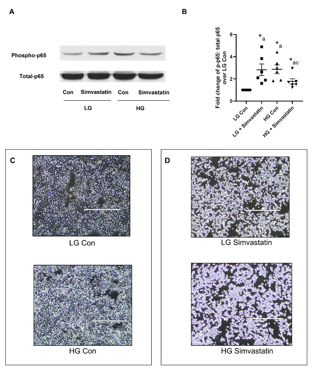
We recently reported a significant increase in S536 phosphorylation of p65 in insulin-secreting INS-1 832/13 cells following exposure to HG conditions [16]. In the current investigation, we undertook a pharmacological approach to further decipher the mechanisms underlying HG-induced phosphorylation of p65. A variety of small molecule inhibitors (Supplementary Table S1) were employed to accomplish our objective. At the outset, we used two structurally distinct inhibitors to assess the roles of protein prenylation in the cascade of events leading to HG-induced phosphorylation of p65. The first is Simvastatin (SMV), a known inhibitor of the cholesterol biosynthetic pathway, which depletes the intracellular pools of mevalonic acid, and its downstream intermediates, such as isoprenyl pyrophosphates that are required for protein prenylation (i.e., farnesylation and geranylgeranylation) [36, 38, 39]. The second inhibitor that we employed is L-788,123, which inhibits G protein (e.g., Rac1) prenylation via inhibition of farnesyl transferase (FTase) and geranylgeranyl transferase (GGTase) [40, 41].
Data depicted in Fig. 1 (Panel A) demonstrate increased phosphorylation of p65 (lane 1 vs . lane 3) in INS-1 832/13 cells following incubation with HG. Co-provision of SMV markedly attenuated HG-induced phosphorylation of p65 (lane 3 vs. lane 4). These data indicate requirement for intermediate(s) of the cholesterol biosynthetic pathway (e.g., isoprenyl pyrophosphates) in HG-induced S536 phosphorylation of p65. Pooled data from six independent studies are graphed in Fig. 1 (Panel B). It is noteworthy that, SMV treatment significantly increased phosphorylation of p65 under LG conditions (lane 1 vs. lane 2), suggesting key roles for intermediates of the cholesterol biosynthetic pathway in retaining p65/RelA in its dephosphorylated state under basal glucose conditions (see below). Furthermore, incubation of INS-1 832/13 cells with SMV, but not the diluent (DMSO) induced clear morphological changes (cell rounding) in these cells (Fig. 1; Panel C and D); these data further validate the hypothesis that protein prenylation plays key roles in cell morphology and cytoskeletal arrangements and architecture [38].
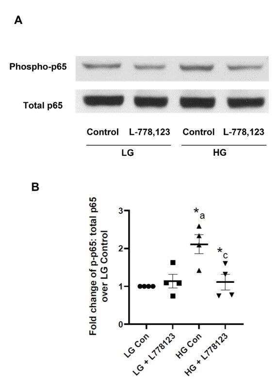
We next assessed the roles of protein prenylation in HG-mediated phosphorylation of p65/RelA by employing L-788,123, a known inhibitor of FTase and GGTase [42, 43]. Data shown in Fig. 2 (Panel A) demonstrate a robust increase in the phosphorylation of p65 under HG exposure conditions (lane 1 vs . lane 3). Co-provision of L-788,123 to the incubation medium markedly suppressed HG-induced phosphorylation of p65 (lane 3 vs . lane 4). Interestingly, unlike SMV, exposure of cells to L-788,123 under LG conditions did not significantly affect the phosphorylation of p65 (lane 1 vs. lane 2). Based on our findings shown in Fig. 1 and 2 we conclude that a protein prenylation-dependent step might be involved in HG-induced S536 phosphorylation of p65. We also propose that increased S536 phosphorylation of p65 seen under LG conditions in the presence of SMV (Fig. 1; lane 1 vs. lane 2) may not be due to a prenylation-dependent step but might be under the regulatory control of other intermediates of the cholesterol biosynthetic pathway. Pooled data from four independent experiments are shown in Fig. 2 (Panel B).
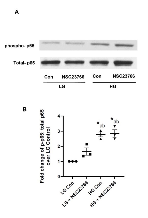
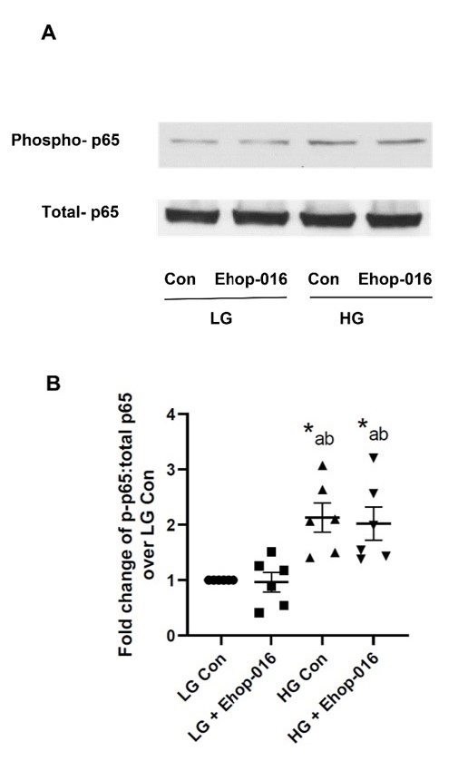
Tiam1-Rac1 and Vav2-Rac1 signaling modules may not contribute to HG-induced p65 phosphorylation in INS-1 832/13 cells
Numerous investigations in a variety of cell types, including retinal endothelial cells [44, 45] and pancreatic beta cells [10, 46-49] have implicated Rac1, a small molecular weight G protein, in metabolic stress-induced cell dysfunction. More importantly, these studies have identified Tiam1 and Vav2 as putative guanine nucleotide exchange factors (GEFs) involved in HG-induced activation of Rac1 [49-51]. In addition, post-translational geranylgeranylation is necessary for appropriate targeting of Rac1 to relevant subcellular compartments for optimal regulation of its effector proteins [39, 47, 52]. In light of our findings that a prenylation-dependent signaling step is necessary for HG-induced phosphorylation of p65, we tested the effects of NSC23766 (inhibitor of Tiam1-Rac1 signaling axis; [48]) and Ehop-016 (inhibitor of Vav2-Rac1 signaling module; [48]) on HG-induced p65 phosphorylation. Data depicted in Fig. 3 (Panel A) demonstrate no significant effects of NSC23766 on phosphorylation of P65 under LG conditions (lane 1 vs. Lane 2). As above, we noted a significant increase in p65 phosphorylation under HG conditions (lane 1 vs. lane 3). Further, HG-induced phosphorylation of p65 was resistant to NSC23766 (lane 3 vs. lane 4). Pooled data from three independent experiments are provided in Fig. 3 (Panel B). Based on these data we conclude that Tiam1-Rac1 signaling step may not be involved in HG-induced p65 phosphorylation.
To assess putative roles of Vav2-Rac1 signaling pathway in HG-induced phosphorylation of p65, we determined the effects of Ehop-016 on HG-induced phosphorylation of p65. Data shown in Fig. 4 (Panel A) demonstrate minimal effects of Ehop-016 on basal (lane 1 vs. lane 2) or HG-induced phosphorylation (lane 3 vs. lane 4) of p65 in INS-1 832/13 cells. Pooled data from six independent studies are included in Fig. 4 (Panel B). Based on the findings in Fig. 3 and 4, we conclude that Rac1 activation mediated by Tiam1 and Vav2 may not mediate HG-induced phosphorylation of p65 in INS-1 832/13 cells.
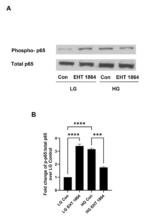
Direct inhibition of Rac1 attenuates p65 phosphorylation under HG-exposure conditions in INS-1 832/13 cells
Next, we sought to further assess the roles of Tiam1/Vav2-independent effects of Rac1 in HG-induced p65 phosphorylation in INS-1 832/13 cells. Désiré and coworkers developed EHT-1864, a small molecular weight compound, which inhibited Rac1 function in vivo [53]. Mechanistic studies have revealed that EHT-1864 binds to Rac1 with high affinity, thereby retaining Rac1 in an inert and inactive state by preventing displacement of pre-bound guanine nucleotides (GDP/GTP) [54, 55]. Earlier studies from our laboratory reported significant inhibition of HG-induced p38MAPK, p53 phosphorylation and cell demise by EHT-1864 in insulin-secreting cells [35, 37, 56]. Therefore, we determined GEF-independent regulatory effects of EHT-1864 on HG-induced phosphorylation of p65 in INS-1 832/13 cells. Data shown in Fig. 5 (Panel A) suggest a significant increase in p65 phosphorylation under basal conditions in the presence of EHT-1864 (lane 1 vs. lane 2). The degree of stimulation by EHT-1864 was comparable to HG-induced effects of p65 phosphorylation (lane 2 vs. lane 3). Interestingly, HG-induced phosphorylation of p65 was significantly inhibited by provision of EHT-1864 (lane 3 vs. lane 4). Pooled data are included in Fig. 5 (Panel B). Based on these data described in Figures 3-5, we conclude that Tiam1-Rac1 and Vav2-Rac1 signaling steps may not underlie HG-induced p65 phosphorylation, and that direct inactivation of Rac1 (by EHT-1864) might exert differential effects on basal and HG-induced phosphorylation of p65. It is also noteworthy that identical effects by SMV (Fig. 1) and EHT-1864 (Fig. 5) were seen on S536 phosphorylation of p65 under LG (stimulation of phosphorylation) and HG (inhibition of phosphorylation) conditions in INS-1 832/13 cells. It is conceivable that SMV elicited effects are mediated via a Rac1-dependent mechanism (see Discussion).
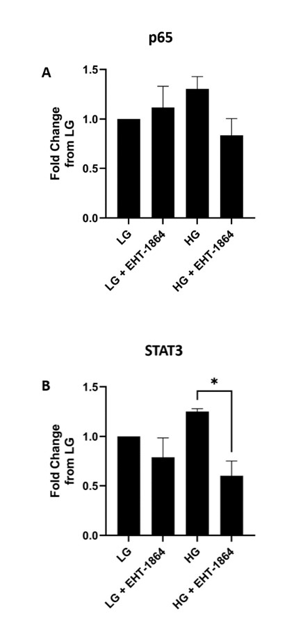
Rac1 activation is necessary for the targeting/association of STAT3, but not p65, with the nuclear fraction in INS-1 832/13 cells under HG exposure conditions
Published evidence in other cell types provide evidence suggesting key regulatory roles for Rho GTPases in the activation of STAT transcription factors [57]. Specifically, studies of Kim and Yoon demonstrated that Rac1 activation is necessary for translocation of NFκB and STAT3 complexes in starved cancer cells [58]. Furthermore, STAT3 has been implicated in beta cell function in health and diabetes [59-61]. Therefore, we undertook a study to quantify relative abundance of total p65 and STAT3 in the nuclear fractions isolated from INS-1 832/13 cells following exposure to LG and HG in the absence and presence of EHT-1864. Data depicted in Fig. 6 indicated no significant effects of Rac1 inhibition on the association of p65 with the nuclear fraction (Panel A). However, EHT-1864 significantly inhibited nuclear accumulation of STAT3 in INS-1 832/13 cells exposed to HG conditions. A modest inhibition of STAT3 association with the nuclear fraction was noted in LG treated cells in the presence of EHT-1864, but such an effect did not reach statistical significance. Taken together, these data suggest that Rac1 might contribute to STAT3 translocation to the nuclear fraction to modulate functions of apoptotic proteins, such as p53 [62], which are known to contribute to HG-induced, Rac1-mediated beta cell dysfunction [35, 37, 63].
CARD9 (Caspase recruitment domain containing protein 9); EHop-016 (N4-(9-ethyl-9H-carbazol-3-yl)-N2-[3-(4-morpholinyl) propyl]-2,4-pyrimidinediamine); EHT-1864 (2-(4-morpholinylmethyl)-5-[[5-[[7-(trifluoromethyl)-4-quinolinyl] thio] pentyl] oxy]-4H-pyran-4-one dihydrochloride); ER stress (endoplasmic reticulum stress); FPP (farnesyl pyrophosphate); FTase (farnesyl transferase); GEFs (guanine nucleotide exchange factors); GGPP (geranylgeranyl pyrophosphate); GGTase (geranylgeranyl transferase); GSIS (glucose-stimulated insulin secretion); GDP/GTP (guanosine diphosphate/guanosine triphosphate); HG (high glucose); IKK (IκB kinase); INGAP (islet neogenesis associated protein); L-778,123 (4-[[5-[[4-(3-chlorophenyl)-3-oxo-1-piperazinyl] methyl]-1H-imidazol-1-yl] methyl]-benzonitrile, monohydrochloride); LG (Low glucose); MSK1 (ribosomal protein S6 kinase alpha-5 kinase); NFκB (nuclear factor κB); Nfr2 (nuclear factor erythroid 2-related factor 2); Nox2 (phagocyte-like NADPH oxidase 2); NSC23766 (N6-[2-[[4-(diethylamino)-1-methylbutyl] amino]-6-methyl-4-pyrimidinyl]-2-methyl-4,6-quinolinediamine, trihydrochloride); P65/RelA (p65 subunit of NFkB); Prx6 (peroxiredoxin 6); Rac1 (ras-related C3 botulinum toxin substrate 1); RSK1 (90 kDa ribosomal S6 kinase); SCF complex (Skp, Cullin, F-box containing complex); SKAP2 (Src kinase-associated phosphoprotein 2); SMV (simvastatin); STAT3 (signal transducer and activator of transcription 3); T1D-T2D (Type 1 diabetes - Type 2 diabetes); TBK1 (TANK-binding kinase 1); Tiam1 (T-lymphoma invasion and metastasis-inducing protein 1); Vav2 (vav guanine nucleotide exchange factor 2).
Author Contributions
SG, MH, NG conducted experiments and analyzed experimental data. AK participated in design of the experiments, overall execution of the studies, and wrote the manuscript.
Funding
This research is supported by a Merit Review Award (BX004663) from the US Department of Veterans Affairs and the National Institutes of Health; EY022230 to AK). AK is the recipient of a Senior Research Career Scientist Award (K6 BX005383) from the US Department of Veterans Affairs. AK would like to thank Wayne State University for Distinguished Professorship award. NG is supported by a T32 predoctoral fellowship from the Detroit Cardiovascular Research Training Center (NIH-2T32HL120822).
The authors declare that no conflicts of interest exist.
| 1 Poitout V, Robertson RP: Glucolipotoxicity: fuel excess and beta-cell dysfunction. Endocr Rev 2008;29:351-366. https://doi.org/10.1210/er.2007-0023 |
||||
| 2 Prentki M, Peyot ML, Masiello P, Madiraju SRM: Nutrient-Induced Metabolic Stress, Adaptation, Detoxification, and Toxicity in the Pancreatic β-Cell. Diabetes 2020;69:279-290. https://doi.org/10.2337/dbi19-0014 |
||||
| 3 Elumalai S, Karunakaran U, Moon JS, Won KC: NADPH Oxidase (NOX) Targeting in Diabetes: A Special Emphasis on Pancreatic β-Cell Dysfunction. Cells 2021;10:1573. https://doi.org/10.3390/cells10071573 |
||||
| 4 Weir GC: Glucolipotoxicity, β-Cells, and Diabetes: The Emperor Has No Clothes. Diabetes 2020;69:273-278. https://doi.org/10.2337/db19-0138 |
||||
| 5 Lytrivi M, Castell AL, Poitout V, Cnop M: Recent Insights Into Mechanisms of β-Cell Lipo- and Glucolipotoxicity in Type 2 Diabetes. J Mol Biol 2020;432:1514-1534. https://doi.org/10.1016/j.jmb.2019.09.016 |
||||
| 6 Mukherjee N, Lin L, Contreras CJ, Templin AT: β-Cell Death in Diabetes: Past Discoveries, Present Understanding, and Potential Future Advances. Metabolites 2021;11:796. https://doi.org/10.3390/metabo11110796 |
||||
| 7 Christensen AA, Gannon M: The Beta Cell in Type 2 Diabetes. Curr Diab Rep 2019;19:81. https://doi.org/10.1007/s11892-019-1196-4 |
||||
| 8 Hasnain SZ, Prins JB, McGuckin MA: Oxidative and endoplasmic reticulum stress in β-cell dysfunction in diabetes. J Mol Endocrinol 2016;56:R33-54. https://doi.org/10.1530/JME-15-0232 |
||||
| 9 Fonseca SG, Urano F, Burcin M, Gromada J: Stress hypERactivation in the β-cell. Islets 2010;2:1-9. https://doi.org/10.4161/isl.2.1.10456 |
||||
| 10 Kowluru A, Kowluru RA: RACking up ceramide-induced islet beta-cell dysfunction. Biochem Pharmacol 2018;154:161-169. https://doi.org/10.1016/j.bcp.2018.04.026 |
||||
| 11 Halban PA, Polonsky KS, Bowden DW, Hawkins MA, Ling C, Mather KJ, Powers AC, Rhodes CJ, Sussel L, Weir GC: β-cell failure in type 2 diabetes: postulated mechanisms and prospects for prevention and treatment. Diabetes Care 2014;37:1751-1758. https://doi.org/10.2337/dc14-0396 |
||||
| 12 Kowluru A, Kowluru RA: Phagocyte-like NADPH oxidase [Nox2] in cellular dysfunction in models of glucolipotoxicity and diabetes. Biochem Pharmacol 2014;88:275-283. https://doi.org/10.1016/j.bcp.2014.01.017 |
||||
| 13 Kowluru A: Oxidative Stress in Cytokine-Induced Dysfunction of the Pancreatic Beta Cell: Known Knowns and Known Unknowns. Metabolites 2020;10:480. https://doi.org/10.3390/metabo10120480 |
||||
| 14 Morgan D, Oliveira-Emilio HR, Keane D, Hirata AE, Santos da Rocha M, Bordin S, Curi R, Newsholme P, Carpinelli AR: Glucose, palmitate and pro-inflammatory cytokines modulate production and activity of a phagocyte-like NADPH oxidase in rat pancreatic islets and a clonal beta cell line. Diabetologia 2007;50:359-369. https://doi.org/10.1007/s00125-006-0462-6 |
||||
| 15 Newsholme P, Morgan D, Rebelato E, Oliveira-Emilio HC, Procopio J, Curi R, Carpinelli A: Insights into the critical role of NADPH oxidase(s) in the normal and dysregulated pancreatic beta cell. Diabetologia 2009;52:2489-2498. https://doi.org/10.1007/s00125-009-1536-z |
||||
| 16 Gamage S, Hali M, Chen F, Kowluru A: CARD9 Mediates Pancreatic Islet Beta-Cell Dysfunction Under the Duress of Hyperglycemic Stress. Cell Physiol Biochem 2022;56:120-137. https://doi.org/10.33594/000000508 |
||||
| 17 Sethi G, Sung B, Aggarwal BB: Nuclear factor-kappaB activation: from bench to bedside. Experimental biology and medicine (Maywood, NJ) 2008;233:21-31. https://doi.org/10.3181/0707-MR-196 |
||||
| 18 Zinatizadeh MR, Schock B, Chalbatani GM, Zarandi PK, Jalali SA, Miri SR: The Nuclear Factor Kappa B (NF-kB) signaling in cancer development and immune diseases. Genes Dis 2021;8:287-297. https://doi.org/10.1016/j.gendis.2020.06.005 |
||||
| 19 Nordmann TM, Dror E, Schulze F, Traub S, Berishvili E, Barbieux C, Böni-Schnetzler M, Donath MY: The Role of Inflammation in β-cell Dedifferentiation. Sci Rep 2017;7:6285. https://doi.org/10.1038/s41598-017-06731-w |
||||
| 20 Hayden MS, Ghosh S: Shared principles in NF-kappaB signaling. Cell 2008;132:344-362. https://doi.org/10.1016/j.cell.2008.01.020 |
||||
| 21 Mitchell S, Vargas J, Hoffmann A: Signaling via the NFκB system. Wiley Interdiscip Rev Syst Biol Med 2016;8:227-241. https://doi.org/10.1002/wsbm.1331 |
||||
| 22 Thoms HC, Stark LA: The NF-κB Nucleolar Stress Response Pathway. Biomedicines 2021;9:1082. https://doi.org/10.3390/biomedicines9091082 |
||||
| 23 Kaltschmidt B, Greiner JFW, Kadhim HM, Kaltschmidt C: Subunit-Specific Role of NF-κB in Cancer. Biomedicines 2018;6:44. https://doi.org/10.3390/biomedicines6020044 |
||||
| 24 Yang B, Maddison LA, Zaborska KE, Dai C, Yin L, Tang Z, Zang L, Jacobson DA, Powers AC, Chen W: RIPK3-mediated inflammation is a conserved β cell response to ER stress. Sci Adv 2020;6:eabd7272. https://doi.org/10.1126/sciadv.abd7272 |
||||
| 25 Carrero JA, Calderon B, Towfic F, Artyomov MN, Unanue ER: Defining the transcriptional and cellular landscape of type 1 diabetes in the NOD mouse. PLoS One 2013;8:e59701. https://doi.org/10.1371/journal.pone.0059701 |
||||
| 26 Veluthakal R, Oh E, Ahn M, Chatterjee Bhowmick D, Thurmond DC: Syntaxin 4 Mediates NF-κB Signaling and Chemokine Ligand Expression via Specific Interaction With IκBβ. Diabetes 2021;70:889-902. https://doi.org/10.2337/db20-0868 |
||||
| 27 Lu X, Yarbrough WG: Negative regulation of RelA phosphorylation: emerging players and their roles in cancer. Cytokine Growth Factor Rev 2015;26:7-13. https://doi.org/10.1016/j.cytogfr.2014.09.003 |
||||
| 28 Sakurai H, Chiba H, Miyoshi H, Sugita T, Toriumi W: IkappaB kinases phosphorylate NF-kappaB p65 subunit on serine 536 in the transactivation domain. J Biol Chem 1999;274:30353-30356. https://doi.org/10.1074/jbc.274.43.30353 |
||||
| 29 Bohuslav J, Chen LF, Kwon H, Mu Y, Greene WC: p53 induces NF-kappaB activation by an IkappaB kinase-independent mechanism involving phosphorylation of p65 by ribosomal S6 kinase 1. J Biol Chem 2004;279:26115-26125. https://doi.org/10.1074/jbc.M313509200 |
||||
| 30 Sakurai H, Suzuki S, Kawasaki N, Nakano H, Okazaki T, Chino A, Doi T, Saiki I: Tumor necrosis factor-alpha-induced IKK phosphorylation of NF-kappaB p65 on serine 536 is mediated through the TRAF2, TRAF5, and TAK1 signaling pathway. J Biol Chem 2003;278:36916-36923. https://doi.org/10.1074/jbc.M301598200 |
||||
| 31 Sasaki CY, Barberi TJ, Ghosh P, Longo DL: Phosphorylation of RelA/p65 on serine 536 defines an I{kappa}B{alpha}-independent NF-{kappa}B pathway. J Biol Chem 2005;280:34538-34547. https://doi.org/10.1074/jbc.M504943200 |
||||
| 32 Schmitz ML, Mattioli I, Buss H, Kracht M: NF-kappaB: a multifaceted transcription factor regulated at several levels. Chembiochem 2004;5:1348-1358. https://doi.org/10.1002/cbic.200400144 |
||||
| 33 Christian F, Smith EL, Carmody RJ: The Regulation of NF-κB Subunits by Phosphorylation. Cells 2016;5:12. https://doi.org/10.3390/cells5010012 |
||||
| 34 Syeda K, Mohammed AM, Arora DK, Kowluru A: Glucotoxic conditions induce endoplasmic reticulum stress to cause caspase 3 mediated lamin B degradation in pancreatic β-cells: protection by nifedipine. Biochem Pharmacol 2013;86:1338-1346. https://doi.org/10.1016/j.bcp.2013.08.023 |
||||
| 35 Sidarala V, Kowluru A: Exposure to chronic hyperglycemic conditions results in Ras-related C3 botulinum toxin substrate 1 (Rac1)-mediated activation of p53 and ATM kinase in pancreatic beta-cells. Apoptosis 2017;22:597-607. https://doi.org/10.1007/s10495-017-1354-6 |
||||
| 36 Baidwan S, Chekuri A, Hynds DL, Kowluru A: Glucotoxicity promotes aberrant activation and mislocalization of Ras-related C3 botulinum toxin substrate 1 [Rac1] and metabolic dysfunction in pancreatic islet β-cells: reversal of such metabolic defects by metformin. Apoptosis 2017;22:1380-1393. https://doi.org/10.1007/s10495-017-1409-8 |
||||
| 37 Sidarala V: Mechanisms of Pancreatic Beta Cell Dysfunction in Diabetes, 2016. PhD Thesis, Wayne State University, Detroit, MI, USA. URL: https://digitalcommons.wayne.edu/oa_dissertations/1591/. | ||||
| 38 Syeda KG, Kowluru A: Inhibition of Prenylation Promotes Caspase 3 Activation, Lamin B Degradation and Loss in Metabolic Cell Viability in Pancreatic β-Cells. Cell Physiol Biochem 2017;43:1052-1063. https://doi.org/10.1159/000481702 |
||||
| 39 Kowluru A, Kowluru RA: Protein prenylation in islet beta-cell function in health and diabetes: Putting the pieces of the puzzle together. Biochem Pharmacol 2015;98:363-370. https://doi.org/10.1016/j.bcp.2015.07.004 |
||||
| 40 Lobell RB, Liu D, Buser CA, Davide JP, DePuy E, Hamilton K, Koblan KS, Lee Y, Mosser S, Motzel SL, Abbruzzese JL, Fuchs CS, Rowinsky EK, Rubin EH, Sharma S, Deutsch PJ, Mazina KE, Morrison BW, Wildonger L, Yao SL, Kohl NE: Preclinical and clinical pharmacodynamic assessment of L-778,123, a dual inhibitor of farnesyl:protein transferase and geranylgeranyl:protein transferase type-I. Mol Cancer Ther 2002;1:747-758. | ||||
| 41 Si MS, Reitz BA, Borie DC: Inhibition of lymphocyte activation and function by the prenylation inhibitor L-778,123. Invest New Drugs 2005;23:21-29. https://doi.org/10.1023/B:DRUG.0000047102.26698.08 |
||||
| 42 Reid TS, Long SB, Beese LS: Crystallographic analysis reveals that anticancer clinical candidate L-778,123 inhibits protein farnesyltransferase and geranylgeranyltransferase-I by different binding modes. Biochemistry 2004;43:9000-9008. https://doi.org/10.1021/bi049280b |
||||
| 43 Marchwicka A, Kamińska D, Monirialamdari M, Błażewska KM, Gendaszewska-Darmach E: Protein Prenyltransferases and Their Inhibitors: Structural and Functional Characterization. Int J Mol Sci 2022;23:5424. https://doi.org/10.3390/ijms23105424 |
||||
| 44 Kowluru RA, Kowluru A, Veluthakal R, Mohammad G, Syed I, Santos JM, Mishra M: TIAM1-RAC1 signalling axis-mediated activation of NADPH oxidase-2 initiates mitochondrial damage in the development of diabetic retinopathy. Diabetologia 2014;57:1047-1056. https://doi.org/10.1007/s00125-014-3194-z |
||||
| 45 Kowluru RA, Mishra M, Kumar B: Diabetic retinopathy and transcriptional regulation of a small molecular weight G-Protein, Rac1. Exp Eye Res 2016;147:72-77. https://doi.org/10.1016/j.exer.2016.04.014 |
||||
| 46 Kowluru A: Friendly, and not so friendly, roles of Rac1 in islet β-cell function: lessons learnt from pharmacological and molecular biological approaches. Biochem Pharmacol 2011;81:965-975. https://doi.org/10.1016/j.bcp.2011.01.013 |
||||
| 47 Kowluru A: Inappropriate movement of Rac1 contributes to glucotoxicity of the islet beta-cell. Cell Cycle 2017;16:1387-1388. https://doi.org/10.1080/15384101.2017.1345229 |
||||
| 48 Kowluru A: Tiam1/Vav2-Rac1 axis: A tug-of-war between islet function and dysfunction. Biochem Pharmacol 2017;132:9-17. https://doi.org/10.1016/j.bcp.2017.02.007 |
||||
| 49 Kowluru A: GPCRs, G Proteins, and Their Impact on β-cell Function. Compr Physiol 2020;10:453-490. https://doi.org/10.1002/cphy.c190028 |
||||
| 50 Kowluru A: Multiple Guanine Nucleotide Exchange Factors Mediate Glucose-Induced Rac1 Activation and Insulin Secretion: Is It Precise Regulatory Control or a Case of Two Peas from the Same Pod? ACS Pharmacol Transl Sci 2021;4:1702-1704. https://doi.org/10.1021/acsptsci.1c00190 |
||||
| 51 Kowluru A: Small G proteins in islet beta-cell function. Endocr Rev 2010;31:52-78. https://doi.org/10.1210/er.2009-0022 |
||||
| 52 Kowluru A: Protein prenylation in glucose-induced insulin secretion from the pancreatic islet beta cell: a perspective. J Cell Mol Med 2008;12:164-173. https://doi.org/10.1111/j.1582-4934.2007.00168.x |
||||
| 53 Désiré L, Bourdin J, Loiseau N, Peillon H, Picard V, De Oliveira C, Bachelot F, Leblond B, Taverne T, Beausoleil E, Lacombe S, Drouin D, Schweighoffer F: RAC1 inhibition targets amyloid precursor protein processing by gamma-secretase and decreases Abeta production in vitro and in vivo . J Biol Chem 2005;280:37516-37525. https://doi.org/10.1074/jbc.M507913200 |
||||
| 54 Shutes A, Onesto C, Picard V, Leblond B, Schweighoffer F, Der CJ: Specificity and mechanism of action of EHT 1864, a novel small molecule inhibitor of Rac family small GTPases. J Biol Chem 2007;282:35666-35678. https://doi.org/10.1074/jbc.M703571200 |
||||
| 55 Onesto C, Shutes A, Picard V, Schweighoffer F, Der CJ: Characterization of EHT 1864, a novel small molecule inhibitor of Rac family small GTPases. Methods Enzymol 2008;439:111-129. https://doi.org/10.1016/S0076-6879(07)00409-0 |
||||
| 56 Sidarala V, Veluthakal R, Syeda K, Vlaar C, Newsholme P, Kowluru A: Phagocyte-like NADPH oxidase (Nox2) promotes activation of p38MAPK in pancreatic beta-cells under glucotoxic conditions: Evidence for a requisite role of Ras-related C3 botulinum toxin substrate 1 (Rac1). Biochem Pharmacol 2015;95:301-310. https://doi.org/10.1016/j.bcp.2015.04.001 |
||||
| 57 Corry J, Mott HR, Owen D: Activation of STAT transcription factors by the Rho-family GTPases. Biochem Soc Trans 2020;48:2213-2227. https://doi.org/10.1042/BST20200468 |
||||
| 58 Kim SJ, Yoon S: Activated Rac1 regulates the degradation of IκBα and the nuclear translocation of STAT3-NFκB complexes in starved cancer cells. Exp Mol Med 2016;48:e231. https://doi.org/10.1038/emm.2016.17 |
||||
| 59 Russell MA, Morgan NG: The impact of anti-inflammatory cytokines on the pancreatic β-cell. Islets 2014;6:e950547. https://doi.org/10.4161/19382014.2014.950547 |
||||
| 60 Weng Q, Zhao M, Zheng J, Yang L, Xu Z, Zhang Z, Wang J, Wang J, Yang B, Richard Lu Q, Ying M, He Q: STAT3 dictates β-cell apoptosis by modulating PTEN in streptozocin-induced hyperglycemia. Cell Death Differ 2020;27:130-145. https://doi.org/10.1038/s41418-019-0344-3 |
||||
| 61 Kostromina E, Wang X, Han W: Altered islet morphology but normal islet secretory function in vitro in a mouse model with microvascular alterations in the pancreas. PLoS One 2013;8:e71277. https://doi.org/10.1371/journal.pone.0071277 |
||||
| 62 Pham TH, Park HM, Kim J, Hong JT, Yoon DY: STAT3 and p53: Dual Target for Cancer Therapy. Biomedicines 2020;8:637. https://doi.org/10.3390/biomedicines8120637 |
||||
| 63 Sidarala V, Kowluru A: The Regulatory Roles of Mitogen-Activated Protein Kinase (MAPK) Pathways in Health and Diabetes: Lessons Learned from the Pancreatic beta-Cell. Recent Pat Endocr Metab Immune Drug Discov 2017;10:76-84. https://doi.org/10.2174/1872214810666161020154905 |
||||
| 64 Yeung YT, Aziz F, Guerrero-Castilla A, Arguelles S: Signaling Pathways in Inflammation and Anti-inflammatory Therapies. Curr Pharm Des 2018;24:1449-1484. https://doi.org/10.2174/1381612824666180327165604 |
||||
| 65 Boyer L, Travaglione S, Falzano L, Gauthier NC, Popoff MR, Lemichez E, Fiorentini C, Fabbri A: Rac GTPase instructs nuclear factor-kappaB activation by conveying the SCF complex and IkBalpha to the ruffling membranes. Mol Biol Cell 2004;15:1124-1133. https://doi.org/10.1091/mbc.e03-05-0301 |
||||
| 66 Sobuz SU, Sato Y, Yoshizawa T, Karim F, Ono K, Sawa T, Miyamoto Y, Oka M, Yamagata K: SIRT7 regulates the nuclear export of NF-κB p65 by deacetylating Ran. Biochim Biophys Acta Mol Cell Res 2019;1866:1355-1367. https://doi.org/10.1016/j.bbamcr.2019.05.001 |
||||
| 67 Ganesan K, Ramkumar KM, Xu B: Vitexin restores pancreatic β-cell function and insulin signaling through Nrf2 and NF-κB signaling pathways. Eur J Pharmacol 2020;888:173606. https://doi.org/10.1016/j.ejphar.2020.173606 |
||||
| 68 Darwish MA, Abo-Youssef AM, Messiha BAS, Abo-Saif AA, Abdel-Bakky MS: Resveratrol inhibits macrophage infiltration of pancreatic islets in streptozotocin-induced type 1 diabetic mice via attenuation of the CXCL16/NF-κΒ p65 signaling pathway. Life Sci 2021;272:119250. https://doi.org/10.1016/j.lfs.2021.119250 |
||||
| 69 Puddu A, Sanguineti R, Durante A, Viviani GL: Pioglitazone attenuates the detrimental effects of advanced glycation end-products in the pancreatic beta cell line HIT-T15. Regul Pept 2012;177:79-84. https://doi.org/10.1016/j.regpep.2012.05.089 |
||||
| 70 Nano E, Petropavlovskaia M, Rosenberg L: Islet neogenesis associated protein (INGAP) protects pancreatic β cells from IL-1β and IFNγ-induced apoptosis. Cell Death Discov 2021;7:56. https://doi.org/10.1038/s41420-021-00441-z |
||||
| 71 Novoselova EG, Glushkova OV, Lunin SM, Khrenov MO, Parfenyuk SB, Novoselova TV, Sharapov MG, Novoselov VI, Fesenko EE: Peroxiredoxin 6 Attenuates Alloxan-Induced Type 1 Diabetes Mellitus in Mice and Cytokine-Induced Cytotoxicity in RIN-m5F Beta Cells. J Diabetes Res 2020;2020:7523892. https://doi.org/10.1155/2020/7523892 |
||||
| 72 Fløyel T, Meyerovich K, Prause MC, Kaur S, Frørup C, Mortensen HB, Nielsen LB, Pociot F, Cardozo AK, Størling J: SKAP2, a Candidate Gene for Type 1 Diabetes, Regulates β-Cell Apoptosis and Glycemic Control in Newly Diagnosed Patients. Diabetes 2021;70:464-476. https://doi.org/10.2337/db20-0092 |
||||
| 73 Sidarala V, Veluthakal R, Syeda K, Kowluru A: EHT 1864, a small molecule inhibitor of Ras-related C3 botulinum toxin substrate 1 (Rac1), attenuates glucose-stimulated insulin secretion in pancreatic beta-cells. Cell Signal 2015;27:1159-1167. https://doi.org/10.1016/j.cellsig.2015.02.020 |
||||
| 74 Kaur S, Mirza AH, Overgaard AJ, Pociot F, Størling J: A Dual Systems Genetics Approach Identifies Common Genes, Networks, and Pathways for Type 1 and 2 Diabetes in Human Islets. Front Genet 2021;12:630109. https://doi.org/10.3389/fgene.2021.630109 |
||||
| 75 Veluthakal R, Tunduguru R, Arora DK, Sidarala V, Syeda K, Vlaar CP, Thurmond DC, Kowluru A: VAV2, a guanine nucleotide exchange factor for Rac1, regulates glucose-stimulated insulin secretion in pancreatic beta cells. Diabetologia 2015;58:2573-2581. https://doi.org/10.1007/s00125-015-3707-4 |
||||