Corresponding Author: Weimin Fan

Department of Orthopedics, the First Affiliated Hospital with Nanjing Medical University, Nanjing, Jiangsu (China)
E-Mail fanweimin@vip.sina.com


Erratum

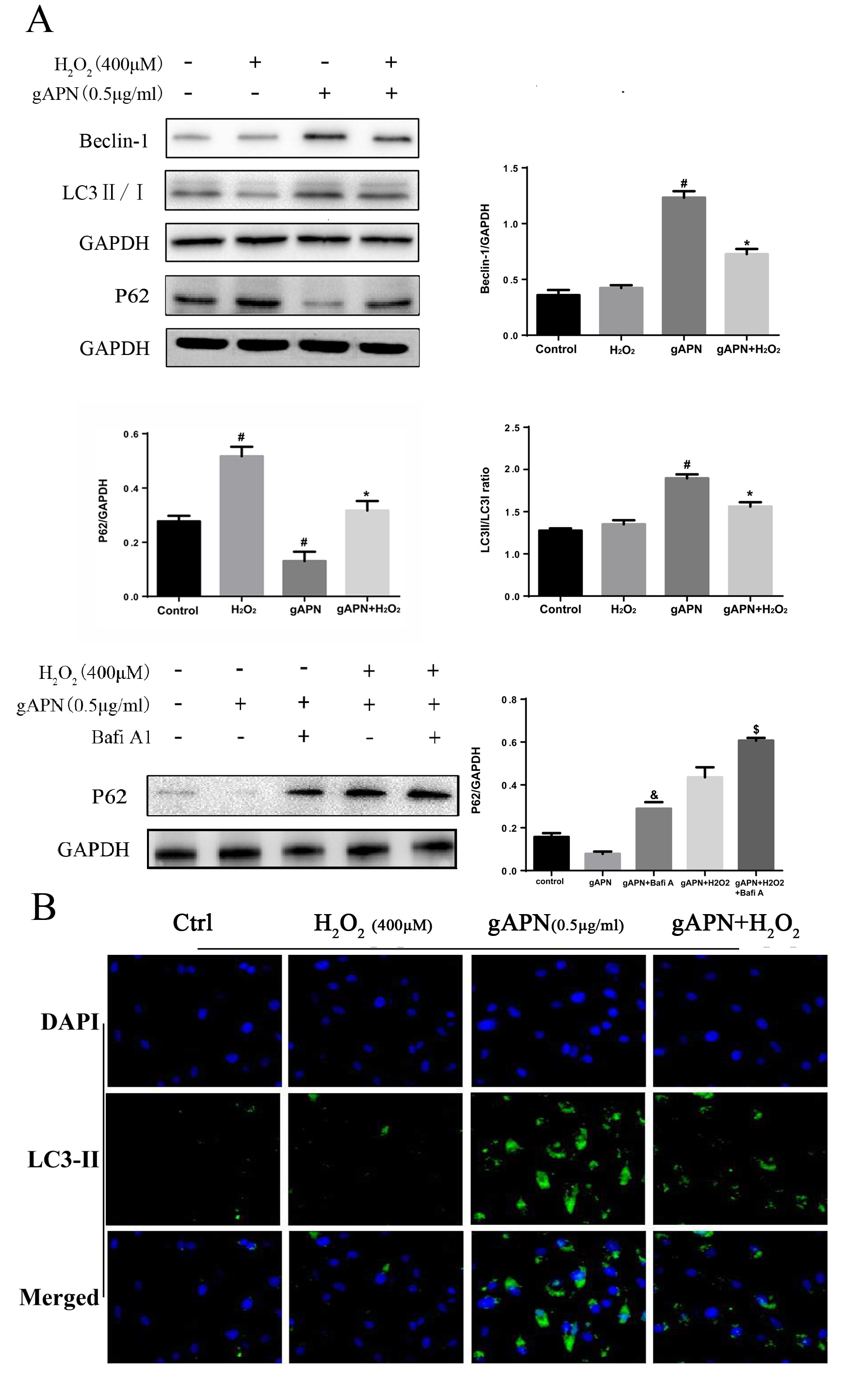
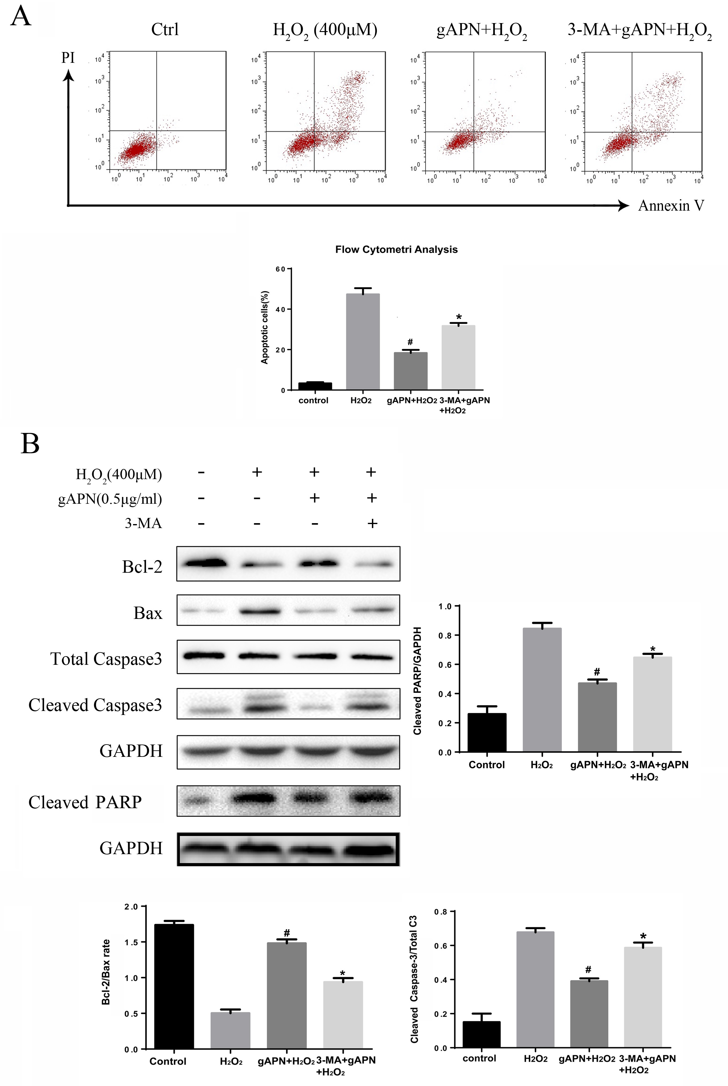
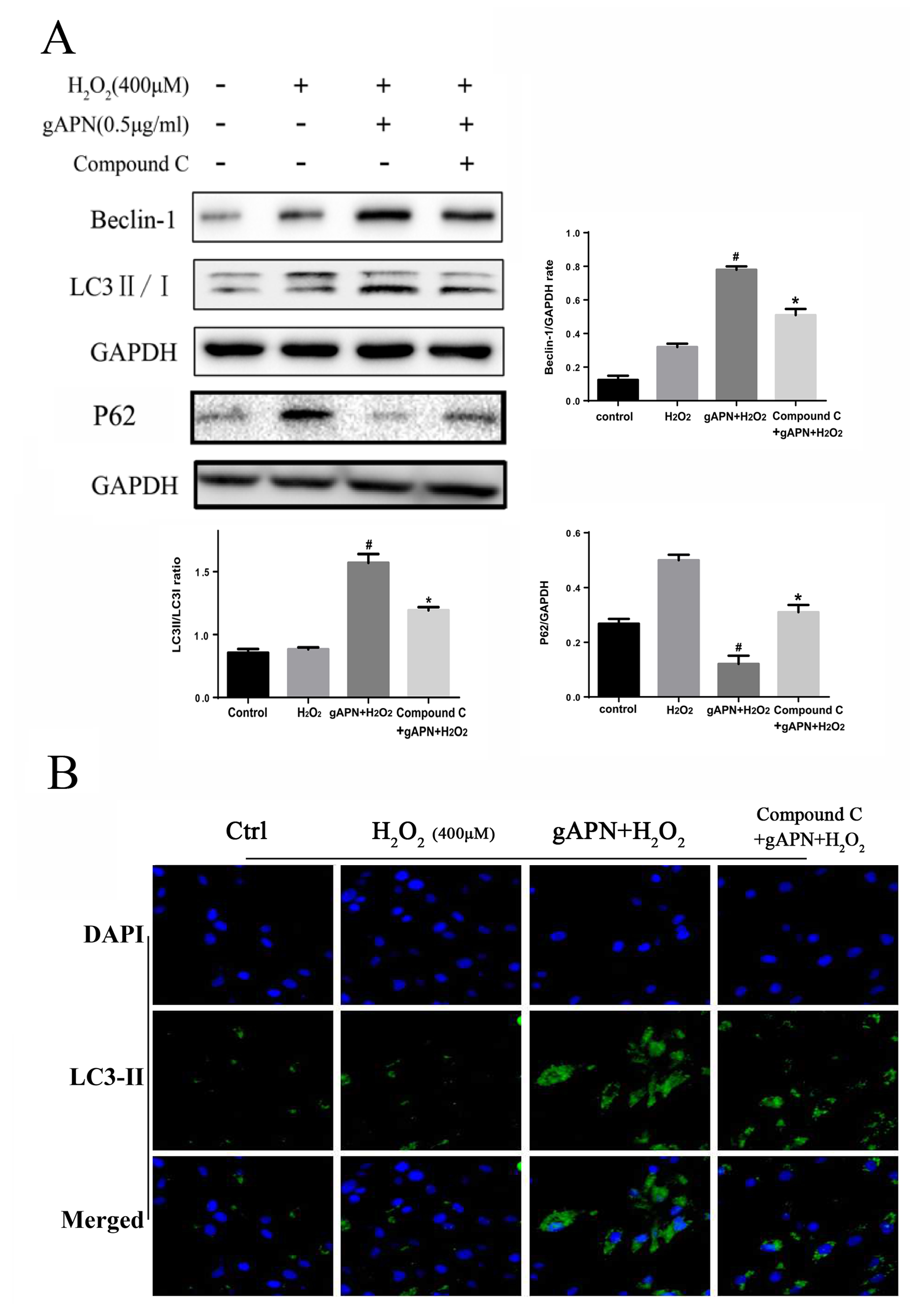
In the article “Globular Adiponectin Attenuated H2O2-Induced Apoptosis in Rat Chondrocytes by Inducing Autophagy Through the AMPK/mTOR Pathway” [Cell Physiol Biochem 2017;43:367- 382, DOI: 10.1159/000480416] by Hu, et al., Figure 7A contains a misplaced band (the band for p62 expression from Figure 4A was repeated), and in Figure 4A, 5B and 7A the images for three bands for the GADPH expression were mismatched.
The authors state that this has been a careless mistake during the preparation of the manuscript due to confusing the respective files because of how they were named.
The authors have provided the data and the corrected figures, which are shown here.

Fig. 4. Effects of H2O2 and gAPN on autophagosome formation in chondrocytes. Chondrocytes were pretreated with 0.5 µg/ mL gAPN for 24 h then treated with or without 400 µM H2O2 for 6 h. (A) Expressions of LC3B, Beclin-1 and P62 by Western blot. (B) LC3-II immunostaining. Significantly increased green bright puncta showed the formation of autophagosome. Results are presented as the ratio of LC3-II to LC3-I and P62,Beclin-1 to GAPDH. Data are represented as the mean±SEM of three independent experiments. #P<0.05 versus the H2O2 group , *&P<0.05 versus the gAPN group and $P<0.05 versus the gAPN+H2O2 group. H2O2, hydrogen peroxide; gAPN, globular adiponectin; BafiA1, Bafilomycin A1, a specific autophagosome-lysosome inhibitor;GAPDH, glyceraldehyde 3-phosphate dehydrogenase.
Fig. 5. Role of autophagy induction by gAPN in the suppression of H2O2-induced apoptosis in chondrocytes. Chondrocytes were treated with 0.5 µg/mL gAPN or pretreated with 10 µM 3-MA for 4 h before gAPN treatment followed by H2O2. (A) Detection of cell apoptosis rate with Annexin V/ PI staining and flow cytometry assays (B) Bcl-2, Bax, Cleaved PARP and Caspase-3 protein expression levels by Western blot. GAPDH was used as a loading control. Results are presented as the ratio of Bcl-2 to Bax and Cleaved caspase-3 to total Caspase-3. Data are represented as the mean ± SEM (n=3). #P<0.05 versus the H2O2 group and *P<0.05 versus the gAPN+H2O2 group. H2O2, hydrogen peroxide; 3-MA, 3-methyl adenine; GAPDH, glyceraldehyde 3-phosphate dehydrogenase.
Fig. 7. Role of AMPK/ mTOR signaling pathway in expression of autophagy-related proteins by gAPN in chondrocytes. Chondrocytes were treated with 0.5 µg/mL gAPN or pretreated with Compound C (10 µM) for 20 min before gAPN treatment prior to H2O2. (A) Expressions of LC3B ,P62 and Beclin-1 after inhibiting the p-AMPK with Compound C by Western blot. (B) Expression of LC3- II after inhibiting the p- AMPK by immunostaining. Results are presented as the ratio of LC3-II to LC3-I and p62, Beclin-1 to GAPDH. Data are represented as the mean ± SEM of three independent experiments. #P<0.05 versus the H2O2. group and *P<0.05 versus the gAPN+ H2O2. group. H2O2, hydrogen peroxide; gAPN, globular adiponectin; Compound C, an inhibitor of AMPK; GAPDH, glyceraldehyde 3-phosphate dehydrogenase.