×
![]()
Corresponding Author: Yunhui Liu
Department of Neurosurgery, Shengjing Hospital of China Medical University,
No. 36, Sanhao Road, Heping District, Shenyang 110004 (China)
E-Mail sj_neurosurgery@126.com
Erratum
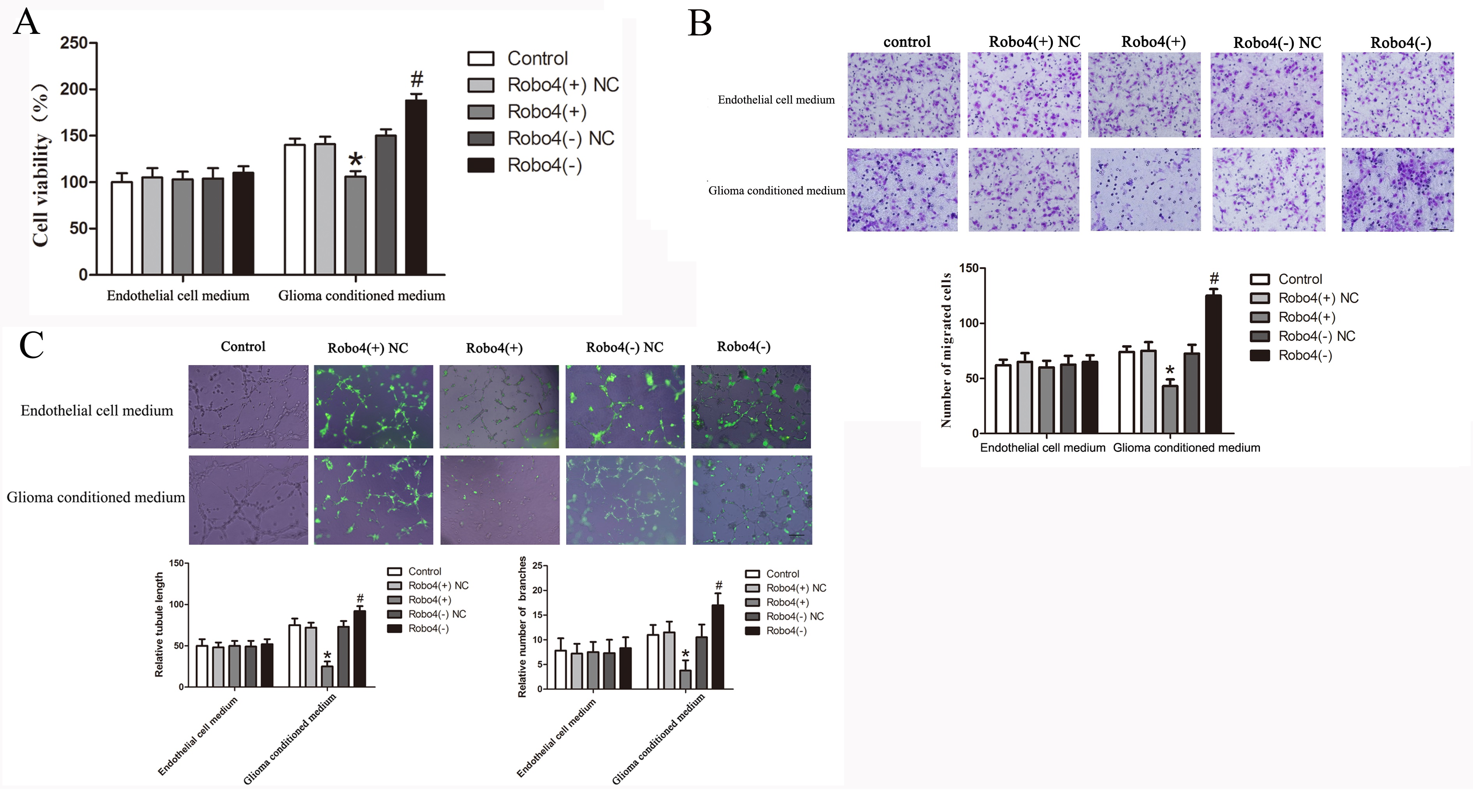
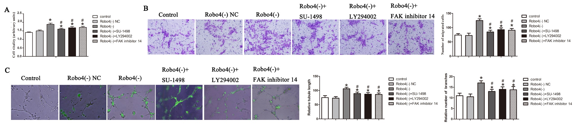
In the article “Roundabout4 Suppresses Glioma-Induced Endothelial Cell Proliferation, Migration
and Tube Formation in Vitro by Inhibiting VEGR2-Mediated PI3K/AKT and FAK Signaling
Pathways” [Cell Physiol Biochem 2015;35:1689-1705. DOI: 10.1159/000373982] by Cai et al.,
a number of incorrect panels were included in Figure 4C and Figure 8C during Figure assembly.
Specifically, Control and Robo4(-) representative images in endothelial cell medium group and
Control, Robo4(-) NC representative images in glioma conditioned medium group of Figure
4C were incorrect in the original article. Control, Robo4(-)NC, Robo4(-), Robo4(-)+LY294002
representative image of Figure 8C was incorrect in the original article.
In preparation of a previous Erratum the authors state that they did not review all the original
images, only those about which concerns were raised. They state that they have now reviewed
all the original data and state that all the original data in this paper are authentic and all the
statistical results and conclusions are correct. The authors submitted the entire original data to
the journal and this submitted data reflects that presented in the figures.
The authors have requested to correct Figure 4C and Figure 8C, due to an error caused by incorrect
assembly of representative pictures in the process of panel incorporation.
The corrected Figure 4 and Figure 8 are shown here.